
Positive Effects of a Brief Session of Aerobic Exercise for Sedentary Children

At a symposium at the 2019 meeting of the American Academy of Child and Adolescent Psychiatry, researcher Benjamin I. Goldstein reported that a single 20-minute session of aerobic exercise (achieving 70% of maximal heart rate) was associated with improvement in cognition and in abnormalities seen on brain imaging in young people. Goldstein urged clinicians to do motivational interviews with sedentary children in their care, emphasizing the positive cardiovascular and cognitive effects of exercise. He indicated this would be more effective than a focus only on weight loss, which is much more difficult to achieve.
Brain Growth in Infancy Predicts Autism

A 2017 article in the journal Nature suggests that brain scans during infancy can predict which kids at risk for autism will go on to develop the disorder, leading to earlier treatment. Studies have shown that children with autism have enlarged brains. The new research zeroes in on the time period when this overgrowth occurs.
Researcher Heather Cody Hazlett and colleagues used magnetic resonance imaging (MRI) scans to measure brain growth in 106 high-risk infants with siblings who have autism spectrum disorder and 42 infants at low risk. The scans were performed when the infants were 6 months, 12 months, and 24 months old.
In 15 infants diagnosed with autism at 24 months, the researchers saw hyperexpansion of cortical surface area between 6 and 12 months and brain overgrowth between 12 and 24 months. The overgrowth coincided with symptoms of autism appearing, and with symptom severity.
The reseachers were able to create a computer algorithm that could predict whether an infant would develop autism based on images of brain growth. The algorithm corrected predicted autism 81% of the time.
Studies have suggested that starting interventions to treat autism early provides the best benefits, so using MRI to diagnose or predict autism before symptoms appear might allow for even earlier treatment that could be more effective.
The study also identified the sites of unusual brain development, which may help researchers determine what mechanisms lead to brain overgrowth in autism and eventually develop treatments that prevent these changes.
Brain Scans Differentiate Suicidal from Non-Suicidal Patients with Bipolar Disorder
 People with bipolar disorder are at high risk for suicidal behavior beginning in adolescence and young adulthood. A 2017 study by Jennifer A. Y. Johnston and colleagues in the American Journal of Psychiatry uses several brain-scanning techniques to identify neurobiological features associated with suicidal behavior in people with bipolar disorder compared to people with bipolar disorder who have never attempted suicide. Clarifying which neural systems are involved in suicidal behavior may allow for better prevention efforts.
The study included 26 participants who had attempted suicide and 42 who had not. Johnston and colleagues used structural, diffusion tensor, and functional magnetic resonance imaging (MRI) techniques to identify differences in the brains of attempters and non-attempters.
Compared to those who had never attempted suicide, those who had exhibited reductions in gray matter volume in the orbitofrontal cortex, hippocampus, and cerebellum. They also had reduced white matter integrity in the uncinate fasciculus, ventral frontal, and right cerebellum regions. In addition, attempters had reduced functional connectivity between the amygdala and the left ventral and right rostral prefrontal cortex. Better right rostral prefrontal connectivity was associated with less suicidal ideation, while better connectivity of the left ventral prefrontal area was linked to less lethal suicide attempts.
Cognitive Behavioral Therapy Improves Depression, PTSD by Improving Brain Connectivity

A recent study clarified how cognitive behavioral therapy improves symptoms of depression and post-traumatic stress disorder (PTSD). The participants were 62 adult women. One group had depression, one had PTSD, and the third was made up of healthy volunteers. None were taking medication at the time of the study. The researchers, led by Yvette Shelive, used functional magnetic resonance imaging (fMRI) to analyze participants’ amygdala connectivity.
At the start of the study, participants with depression or PTSD showed diminished connectivity between the amygdala and brain areas related to cognitive control, the process by which the brain can vary behavior and how it processes information in the moment based on current goals. The lack of connectivity reflected the severity of the participants’ depression. Twelve weeks of cognitive behavioral therapy improved mood and connectivity between the amygdala and these control regions, including the dorsolateral prefrontal cortex and the inferior frontal cortex. These regions also allow for executive functioning, which includes planning, implementation, and focus.
Brain Inflammation in People at High Risk for Schizophrenia
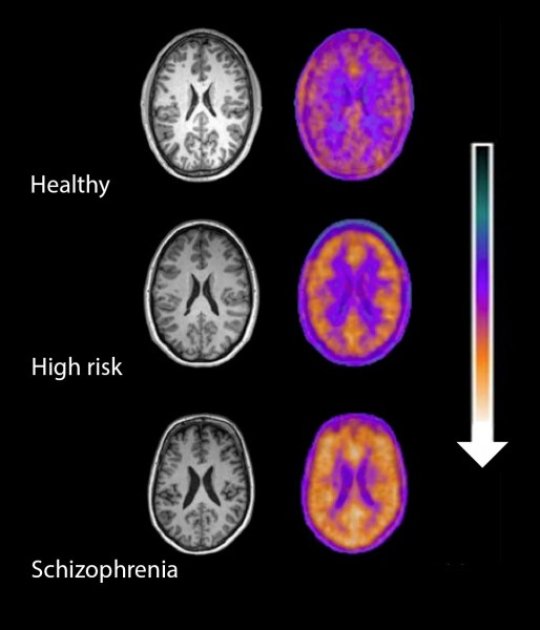
Microglial activity in the brains of people who are healthy, people at high risk for schizophrenia, and people who have been diagnosed with schizophrenia.
A 2016 study by Peter S. Bloomfield and colleagues in the American Journal of Psychiatry used PET scans to compare the activity of microglia, immune cells in the central nervous system, in healthy controls, people with schizophrenia, and those at high risk for the illness. It found that both people with schizophrenia and those at high risk had greater brain inflammation than the healthy controls.
The study was the first to show that microglial activity was elevated in people at high risk (who showed some preliminary symptoms of schizophrenia). The finding had a large effect size.
Microglial activity was also correlated with symptom severity in the high-risk participants. Increased microglial activity was not linked to depression, suggesting that it is specific to the development of psychosis.
These findings resemble those of other recent studies showing increased inflammation in people at high risk for psychosis.
The study suggests that increased microglial activity occurs before a first episode of psychosis. That means it could help identify people who may develop schizophrenia. The findings also suggest that anti-inflammatory treatment could theoretically be used to prevent psychosis.
Lighting Up Neural Networks in Mice
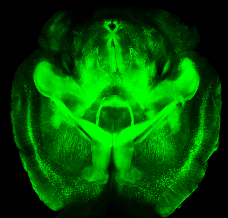 A new technology is making it possible to view the mammalian brain’s structure and connectivity for the first time. Karl Deisseroth discussed the technology, called CLARITY, at a plenary lecture at the 2014 meeting of the International College of Neuropsychopharmacology.
The way CLARITY works is by replacing lipids in the brain with a hydrogel substance. This preserves the structure of the brain’s neural networks, leaves proteins and nucleic acids intact, but allows for observation by rendering the brain transparent. This can be done in a system as large as the entire adult mouse brain. Early attempts took a whole day, but Deisseroth eventually found a way to render a mouse’s brain transparent in a matter of minutes.
The pictures are truly amazing, allowing for the visualization of previously microscropic neurons, dendrites, axons and connections in life-sized images. Pictures and details are available at www.clarityresourcecenter.org.
Deisseroth and colleagues have used CLARITY imaging to determine where neurons fire during different social activities. By placing photosensitive fibers in selected neurons using a virally based gene insertion technique, Deisseroth and colleagues were able to selectively fire dopamine neurons in the ventral tegmental area, part of the brain’s reward system, and thus increase or decrease the social interaction of mice by increasing or decreasing firing. The effects were selective to social interaction; the firing did not affect locomotor activity or exploration of an inanimate object.
The ventral tegmental area contains neurons that project to several locations in the brain, and Deisseroth and colleagues hoped to observe which were important to social interaction. Stimulating the ventral tegmental area to drive the medial prefrontal cortex caused anxiety in the mice and made them averse to social interaction. However, when the ventral tegmental area was used to selectively drive the nucleus accumbens, another part of the brain’s reward system, social interaction increased.
Deisseroth wanted to know if the nucleus accumbens was also involved in normal spontaneous social interactions. The researchers used a virus to insert an opsin-sensitive calcium gene that could give an ongoing readout of neural activity. (Opsin is a light-sensitive receptor found in cells in the retina.) The team found that the nucleus accumbens was implicated in social interaction with another mouse, but not in exploration of a novel object. Based on CLARITY imaging of the structure of ion channels (which are so small they cannot even be seen with an electron microscope), Deisseroth was able to selectively alter ion fluxes and turn neuronal firing on or off at will.
In the last 50 years, the brain and its billions of neurons and hundreds of trillions of synapses have gone from complete inaccessibility toward increasing clarity.
