Brain Imaging Finds Abnormalities that Appear Over the Course of Childhood-Onset Bipolar Illness
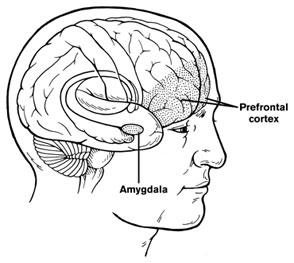
 There is considerable evidence that children with bipolar disorder have smaller amygdalas, and the amygdala also appears to be hyper-reactive when these children perform facial emotion recognition tasks. A symposium on longitudinal imaging studies in pediatric bipolar disorder was held at the 2012 meeting of the American Academy of Child and Adolescent Psychiatry to shed light on other brain abnormalities in these children.
Researcher Nancy Aldeman reported that there is some evidence children with bipolar disorder have decreased gray matter volume in parts of the brain including the subgenual cingulate gyrus, the orbital frontal cortex, and the superior temporal gyrus, as well as the left dorsolateral prefrontal cortex and amygdala. At the same time there is evidence of increased size of the basal ganglia. These abnormalities do not appear to precede the onset of the illness.
Some changes occur over the course of the illness. The basal ganglia seem to increase in volume in patients with bipolar disorder, but decrease in volume in those with severe mood dysregulation and comorbid ADHD. Moreover, parietal cortex and precuneus cortex volumes appeared to increase in children with bipolar disorder while decreasing or staying the same in normal volunteer controls.
A meta-analysis of brain imaging studies indicated that in general, the size of the amygdala appears to increase from childhood to adulthood in bipolar patients, starting out smaller than that of similarly-aged normal volunteers, but becoming larger than that of adult normal volunteers as the patients age into adulthood.
Lithium treatment increases gray matter volume in a variety of cortical areas and in the hippocampus in multiple studies. In contrast, treatment with valproate for 6 weeks appears to decrease hippocampal volume.
Heading Off Early Symptoms of Bipolar Disorder in Children at High Risk
 At the American Academy of Child and Adolescent Psychiatry (AACAP) annual meeting in Toronto in October 2011, there was a symposium on risk and resilience factors in the onset of bipolar disorder in children who have a parent with the disorder.
Family Focused Therapy Highly Encouraged
Amy Garrett reported that family focused therapy (FFT) in those at risk for bipolar disorder was effective in ameliorating symptomatology compared to treatment as usual. Family focused therapy, pioneered by Dave Miklowitz, PhD of UCLA involves three components. The first component is education about the illness and methods of self-management. The second is enhancement of communication in the family with practice and rehearsal of new modes of conversation. The third component is assistance with problem solving.
In Garrett’s study, 50 children aged 7 to 17 were randomized to family focused treatment or treatment as usual. These children were not only at high risk for bipolar disorder, they were already prodromal, meaning they were already diagnosable with bipolar not otherwise specified (BP-NOS), cyclothymia, or major depressive disorder, and had also shown concurrent depressive and/or manic symptoms in the two weeks prior to the study. At baseline, compared to controls, these children at high risk for full-blown bipolar disorder by virtue of a parental history of the illness showed increased activation of the amygdala and decreased activation of the prefrontal cortex. Most interestingly, after improvement with the family focused therapy (FFT), amygdala reactivity to emotional faces became less prominent and dorsolateral prefrontal cortical activity increased in proportion to the degree of the patient’s improvement.
The discussant for the symposium was Kiki Chang of Stanford University, who indicated that the results of this study of family focused therapy were already sufficient to convince him that FFT was a useful therapeutic procedure in children at high risk for bipolar disorder by virtue of having a parent with a history of bipolar illness. Chang is now employing the therapy routinely in all of his high-risk patients.
Editors Note: This is an extremely important recommendation as it gives families a specific therapeutic process in which to engage children and others in the family when affective behavior begins to become abnormal, even if it does not meet full criteria for a bipolar I or bipolar II disorder.
FFT also meets all the important criteria needed for putting it into widespread clinical practice. Family focused therapy has repeatedly been shown to be effective in adults and adolescents with bipolar illness and now also in these children who are prodromal. The psychoeducational part of FFT is common sense, and dealing with communication difficulties and assisting with problem solving also have merit in terms of stress reduction. Finally, this treatment intervention appears to be not only safe but also highly effective in a variety of different prodromal presentations of affect disorders even if children do not meet full criteria for bipolar disorder. While the few studies of early intervention with psychopharmacological agents have not yet identified efficacious medications for the prodromes of bipolar disorder and in particular medications with a high degree of safety, such family focused therapy appears to be an ideal early intervention.
I would concur with Dr. Chang’s assessment. Family focused therapy (FFT) should be offered to all children with this high-risk status who have begun to be symptomatic. Early onset of unipolar depressive disorder or of bipolar disorder carries a more adverse prognosis than the adult onset variety and thus should not be ignored. If more serious illness is headed off early, it even raises the possibility that the full-blown illness will not develop at all.
Gray Matter Volume Abnormalities
Tomas Hajek of Dalhousie University in Halifax presented data indicating that in children at high risk for bipolar disorder, gray matter volume in the right inferior frontal gyrus is increased. Read more
Cortex Shrinks and Amygdala Grows in Childhood Bipolar Disorder
 At a symposium on new research on juvenile bipolar disorder at the meeting of the American Academy of Child and Adolescent Psychiatry (AACAP) in 2010, the discussant Kiki Chang of Stanford University reported some recent neurobiological findings on childhood bipolar disorder. He found evidence that prefrontal cortical volume appears to decrease over the course of the illness and, conversely, there was evidence of increases in amygdala volume. He also found that the volume of the striatum (or caudate nucleus, which is involved in motor control) increased in children with bipolar illness or bipolar illness comorbid with ADHD, but decreased in children with ADHD alone.
Chang cited the study of Singh et al. (2010) who found that the subgenual anterior cingulate volume early in the course of illness was smaller in adolescent-onset bipolar disorder compared to controls. Given this evidence of prefrontal cortical and anterior cingulate deficits, Dr. Chang raised the possibility that treatment with lithium and other agents with potential neurotrophic and neuroprotective effects might be able to prevent these neurobiological aspects of illness progression in young patients.
Thalamic Volume and Neural Connectivity in Autism
 Ish Bhalla reported at the 57th Annual Meeting of the American Academy of Child and Adolescent Psychiatry (AACAP) in October 2010 that children with autism have greater thalamic volume than normal controls. Other posters presented at the meeting showed that patients with autism spectrum disorders have connectivity abnormalities, with increased connectivity of neurons to other nearby neuronal groups and decreased connectivity of neurons to areas of the brain at a greater distance.
Editor’s Note: These findings echo reports that the corpus callosum, the main structure connecting neurons across the two hemispheres of the brain, is smaller in autism. In addition, other investigators have reported abnormalities in cortical column structure in autism.
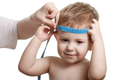
Interestingly, the findings of increased volume and altered connectivity may even be reflected in measurements of brain and head size. A substantial literature supports the observations that children with autism have greater initial head size and growth of their heads compared with the normal infant population.
Maternal Depression May Affect Child’s Brain
At the 57th Annual Meeting of the American Academy of Child and Adolescent Psychiatry (AACAP) in October 2010, Dana Serino of Columbia University reported that mothers who experienced depression while pregnant had children with increased size in both their left and right medial temporal gyri, parts of the brain that may be responsible for judging distance, recognizing faces, and understanding word meaning.
Editor’s Note: These data extend preclinical studies that have shown that a variety of prenatal stressors are capable of exerting substantial effects on biology and behavior in children. In addition to affecting medial temporal gyrus volume, prenatal depression has also been shown to have effects on behavior of newborns, indicating that depression during pregnancy may have adverse effects on both the mother and the newborn.
New Developments in Repeated Transcranial Magnetic Stimulation (rTMS)
 At the 65th Annual Scientific Convention of the Society of Biological Psychiatry, several findings related to repeated transcranial magnetic stimulation (rTMS) were reported.
E. Baron Short reported that two weeks of 10 Hz rTMS at 120% of motor threshold (MT) was highly effective in the treatment of fibromyalgia. Pain ratings decreased 45% by day six and 80% by day 10 in this randomized sham-controlled double-blind study.
Also at the convention, Motoaki Nakamura reported that either 1 Hz or 20 Hz rTMS at 90-100% of motor threshold over left prefrontal cortex in depressed patients increased gray matter in left dorsolateral prefrontal cortex and left hippocampus in association with almost 50% reductions in Hamilton depression rating scale scores and associated increases in performance on the Wisconsin card sort test. Read more
Brain Volume Reduced As Early As First Episode Of Mania
 Researchers Manpreet K. Singh and Kiki D. Chang et al. from Stanford reported at the 65th Annual Scientific Convention of the Society of Biological Psychiatry that adolescents experiencing a first episode of mania show reduced volume in the subgenual anterior cingulate cortex (Brodmann area 25). Previous studies have indicated that teens and adults with bipolar disorder exhibit decreased volume in prefrontal gray matter. Read more


