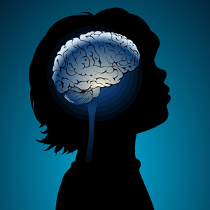
Brain Imaging Finds Abnormalities that Appear Over the Course of Childhood-Onset Bipolar Illness
 There is considerable evidence that children with bipolar disorder have smaller amygdalas, and the amygdala also appears to be hyper-reactive when these children perform facial emotion recognition tasks. A symposium on longitudinal imaging studies in pediatric bipolar disorder was held at the 2012 meeting of the American Academy of Child and Adolescent Psychiatry to shed light on other brain abnormalities in these children.
Researcher Nancy Aldeman reported that there is some evidence children with bipolar disorder have decreased gray matter volume in parts of the brain including the subgenual cingulate gyrus, the orbital frontal cortex, and the superior temporal gyrus, as well as the left dorsolateral prefrontal cortex and amygdala. At the same time there is evidence of increased size of the basal ganglia. These abnormalities do not appear to precede the onset of the illness.
Some changes occur over the course of the illness. The basal ganglia seem to increase in volume in patients with bipolar disorder, but decrease in volume in those with severe mood dysregulation and comorbid ADHD. Moreover, parietal cortex and precuneus cortex volumes appeared to increase in children with bipolar disorder while decreasing or staying the same in normal volunteer controls.
A meta-analysis of brain imaging studies indicated that in general, the size of the amygdala appears to increase from childhood to adulthood in bipolar patients, starting out smaller than that of similarly-aged normal volunteers, but becoming larger than that of adult normal volunteers as the patients age into adulthood.
Lithium treatment increases gray matter volume in a variety of cortical areas and in the hippocampus in multiple studies. In contrast, treatment with valproate for 6 weeks appears to decrease hippocampal volume.

