Brain Growth in Infancy Predicts Autism
A 2017 article in the journal Nature suggests that brain scans during infancy can predict which kids at risk for autism will go on to develop the disorder, leading to earlier treatment. Studies have shown that children with autism have enlarged brains. The new research zeroes in on the time period when this overgrowth occurs.
Researcher Heather Cody Hazlett and colleagues used magnetic resonance imaging (MRI) scans to measure brain growth in 106 high-risk infants with siblings who have autism spectrum disorder and 42 infants at low risk. The scans were performed when the infants were 6 months, 12 months, and 24 months old.
In 15 infants diagnosed with autism at 24 months, the researchers saw hyperexpansion of cortical surface area between 6 and 12 months and brain overgrowth between 12 and 24 months. The overgrowth coincided with symptoms of autism appearing, and with symptom severity.
The reseachers were able to create a computer algorithm that could predict whether an infant would develop autism based on images of brain growth. The algorithm corrected predicted autism 81% of the time.
Studies have suggested that starting interventions to treat autism early provides the best benefits, so using MRI to diagnose or predict autism before symptoms appear might allow for even earlier treatment that could be more effective.
The study also identified the sites of unusual brain development, which may help researchers determine what mechanisms lead to brain overgrowth in autism and eventually develop treatments that prevent these changes.
Diuretic Looks Promising for Autism
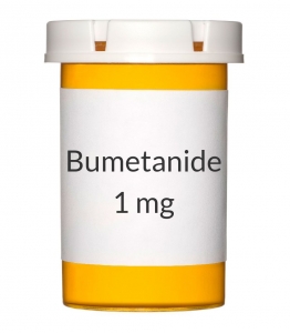
 Phase 2 clinical trials showed that the diuretic bumetanide can reduce the severity of autism spectrum disorders in children aged 3 to 11. A 2017 phase 2B trial assessed side effects and determined the dosage that maximizes benefits and minimizes side effects. Bumetanide will now move on to year-long phase 3 trials in five European countries and may be on the market by late 2021. Bumetanide is an unusually potent ‘loop diuretic’ (a diuretic that works at the loop of Henle in the kidney). In preliminary studies, it has also been used to prevent seizures in newborns.
The phase 2B study included 88 mostly male participants with autism spectrum disorder between the ages of 2 and 18. The participants were randomly assigned to receive 0.5 mg, 1.0 mg, or 2.0 mg twice daily of bumetanide or a placebo for three months.
Bumetanide improved core symptoms of autism such as social communication and restricted interest across all ages. Side effects were worse at higher doses, and included hypokalemia (low potassium), increased urine production, loss of appetite, dehydration, and weakness or lack of energy.
Researchers led by Eric Lemonnier determined that doses of 1.0 mg twice/day produces the most benefits while controlling side effects.
Study of Baby Teeth Links Autism and Exposure to Heavy Metals Such as Lead
Recent research has revealed that autism is linked to new onset genetic mutations (called ‘de novo’ mutations) that occur during early fetal development. A new study suggests that levels of heavy metals such as lead and zinc (but not mercury) may affect the likelihood that these mutations will occur.
The 2017 study by Manish Arora and colleagues in the journal Nature Communications included twins with and without autism, particularly twin pairs in which one twin had autism and the other did not. An international team of scientists collected naturally shed baby teeth from the twins. The researchers then used lasers to extract specific layers of dentine, the hard substance beneath tooth enamel, which correspond to different developmental periods, including before birth and in early childhood. The researchers then analyzed these dentine samples to determine the children’s uptake of various heavy metals in early life.
The analysis showed that children with autism had higher levels of lead (a neurotoxin) throughout development, but particularly right after birth. Children with autism also had lower uptake of manganese, an essential nutrient. Compared to children without autism, children with autism had lower zinc levels in utero, but higher zinc levels after birth. Zinc is another essential nutrient. Lead and manganese levels were also linked to autism severity.
The method of analyzing teeth allows researchers to look back in time and measure what children were exposed to years earlier. This may help identify environmental factors that contribute to autism spectrum disorders.
Autism Linked to Banned Chemicals
 An explanation for the increase in autism rates over the past few decades has remained elusive in the years since researcher Andrew Wakefield fabricated a link between the disorder and mercury in vaccinations that was eventually completely debunked.
In 2016, researcher Kristin Lyall of Drexel University’s A.J. Drexel Autism Institute published findings suggesting that high exposure during pregnancy to chemicals banned in the 1970s increased risk of an autism spectrum disorder.
The study looked at 1144 children born in southern California between 200 and 2003. Their mothers had participated in California’s Expanded Alphafetoprotein Prenatal Screening Program, intended to identify birth defects during pregnancy. Second trimester blood samples from these women could be used to determine to what extent their children were exposed to the chemicals while in utero. The researchers found an association between the highest exposure levels and later autism diagnoses.
Lyall and colleagues measured levels of two different classes of organochlorine chemicals: polychlorinated biphenyls (PCBs), used as lubricants, coolants, and insulators; and organochlorine pesticides (OCPs), including DDT, which was banned in 1972. All production of organochlorine chemicals was banned in the US in 1977, but they remain in the environment and are absorbed in the fat of animals that humans eat. According to Lyall, people in the US generally have detectable levels of organochlorine chemicals in their bodies.
The study revealed that exposure to two compounds in particular—PCB 138/158 and PCB 153—was linked to dramatically higher autism rates. Level of exposure is key to autism risk. Those children in the top 25 percentile of exposure were 79% and 82% more likely to have an autism diagnosis than those with the lowest levels of exposure, respectively.
High exposure to two other compounds, PCB 170 and PCB 180, increased autism risk by 50%.
The findings by Lyall and colleagues were published in the journal Environmental Health Perspectives.
Editor’s Note: Another study by Manish Arora and colleagues links autism risk to levels of lead, zinc and manganese absorbed in early life.
The myth that mercury in vaccines causes autism still lingers in our popular culture. Mercury is no longer used in vaccines, but autism rates are still increasing. Perhaps the new findings of a link between heavy metals and autism will help end the misinformation about the safety of vaccines and allow more parents to vaccinate their children without worry.
Dietary Supplements for Autism: Up-to-Date Research
A 2017 review article by Yong-Jiang Li and colleagues in the journal Frontiers in Psychiatry describes the current research on dietary supplements that may help improve symptoms of autism spectrum disorder.
Some of the most promising research was on vitamin D, folinic acid, and sulforaphane. Methyl B12 and digestive enzyme therapy had some positive effects, while gluten- and casein-free diets and omega-3 fatty acids did not seem to help improve autism symptoms.
Vitamin D
Li and colleagues described a randomized, controlled trial of vitamin D in 109 children with autism aged 3 to 10 years. The experimental group received doses of 300 IU/kg of body weight/day, not exceeding 5000 IU/day. By the end of the four-month study, vitamin D levels had significantly increased in the experimental group compared to the control group. Those who received vitamin D also showed significant improvement on all ratings of autism symptoms, which included general scales of autism symptoms and more specialized checklists that capture aberrant behavior and social responsiveness.
Folinic Acid
The review article also described a randomized double-blind placebo-controlled trial of folinic acid in 48 children with autism spectrum disorder and language impairment. Participants received high-dose folinic acid (2 mg/kg/day) or placebo for 12 weeks. Those who received folinic acid, a form of folic acid that can readily be used by the body, showed significant improvements in verbal communication and core autism symptoms compared to those who received placebo. Participants who tested positive for folate receptor alpha autoantibodies (FRAA), which disrupt the transportation of folate across the blood-brain barrier and are common in autism, showed greater improvements from taking folinic acid than those without this abnormality.
Sulforaphane
Sulforaphane is a phytochemical derived from cruciferous vegetables. It can create metabolic effects that resemble those of a fever, which can improve behavioral symptoms of autism. Sulforaphane also fights oxidative stress, inflammation, and DNA damage, which may play roles in autism. Li and colleagues described the first double-blind, placebo-controlled trial of sulforaphane treatment in 29 boys aged 13 to 17 years. The boys who received sulforaphane showed significant improvement in autism-related behavior, especially social interaction and communication, after 18 weeks compared to those who received placebo. Sulforaphane has low toxicity and is well tolerated. Read more
Successful Trial of N-Acetylcysteine for Veterans with PTSD and Substance Abuse
 The antioxidant N-acetylcysteine (NAC) can improve a number of habit-related conditions, such as substance use disorders, gambling, and compulsive hair-pulling. It also aids in the treatment of depression and obsessive-compulsive disorder (OCD). A 2016 study by Susie E. Back and colleagues in the Journal of Clinical Psychiatry found that NAC can also improve symptoms of post-traumatic stress disorder (PTSD) in veterans who also had substance use disorders.
In the pilot study of 35 veterans, participants were randomized to receive an 8-week course of NAC (2,400 mg/day) or placebo, plus cognitive-behavioral therapy targeting their substance use disorder. PTSD and substance use disorders have some overlapping neurobiological features, such as impaired prefrontal cortex regulation of basal ganglia circuitry.
At the end of the 8-week trial, those veterans who received NAC showed improvement in PTSD symptoms, substance cravings, and depression compared to those who received placebo. Substance use was similar and low among both groups. Side effects were minimal.
While these results were preliminary, they suggest that NAC could treat both PTSD and substance use disorders, which often occur together. Larger studies are expected to follow.
Editor’s Note: These preliminary data add to the evidence that NAC has remarkably wide utility in addictions (cocaine, alcohol, nicotine, and marijuana), habits (including OCD, trichotillomania/hair-pulling, nail biting, skin-picking, and cutting), depression and anxiety in bipolar disorder and negative symptoms in schizophrenia.
Different Types of Trauma Affect Brain Volume Differently
Post-traumatic stress disorder (PTSD) has been associated with decreased volume of gray matter in the cortex. Research by Linghui Meng and colleagues has revealed that the specific types of trauma that precede PTSD affect gray matter volume differently.
At the 2016 meeting of the Society for Neuroscience, Meng reported that PTSD from accidents, natural disasters, and combat led to different patterns of gray matter loss. PTSD from accidents was associated with gray matter reductions in the bilateral anterior cingulate cortex (ACC) and medial prefrontal cortex (mPFC). PTSD from natural disasters was linked to gray matter reductions in the mPFC and ACC, plus the amygdala and left hippocampus. PTSD from combat reduced gray matter volume in the left striatum, the left insula, and the left middle temporal gyrus.
Meng and colleagues also found that severity of PTSD was linked to the severity of gray matter reductions in the bilateral ACC and the mPFC.
In a 2016 article in the journal Scientific Reports, Meng and colleagues reported that single-incident traumas were associated with gray matter loss in the bilateral mPFC, the ACC, insula, striatum, left hippocampus, and the amygdala, while prolonged or recurrent traumas were linked to gray matter loss in the left insula, striatum, amygdala, and middle temporal gyrus.
Steroid Dexamethasone Facilitates Fear Extinction
 A 2017 article by Vasiliki Michopoulos and colleagues in the journal Psychoendocrinology reports that the potent steroid dexamethasone reduced the fear-potentiated startle response in patients with post-traumatic stress disorder (PTSD) but not in healthy controls. Dexamethasone acts on glucocorticoid receptors to suppress the body’s secretion of cortisol.
In the study, participants both with and without PTSD were taught to associate a picture of a blue square or a purple triangle with an uncomfortable short blast of air to the larynx (voicebox) and a loud burst of broadband noise in the participants’ headphones.
Some participants were given a placebo the night before the study, while others received a 0.5 mg dose of dexamethasone. Those who received dexamethasone the night before the study acquired a startle response to the blue square or purple triangle as much as other participants.
People without PTSD were easily able to eliminate their fear of the visual symbol when it was no longer linked to the noise and the blast of air, regardless of whether they had taken dexamethasone. However, among those with PTSD, only those who received dexamethasone were able to eliminate this fear-potentiated startle response and properly discriminate between safe and unsafe signals. People with PTSD who received the placebo maintained the fearful response to the blue square or purple triangle and startled in response to safe symbols.
People with PTSD may have difficulty learning to inhibit their fearful responses to stimuli that are no longer dangerous. In this study, the patients with PTSD continued to startle even after repeated presentations of the visual symbol without any accompanying air blast, while the controls showed excellent extinction of the response. After dexamethasone, but not placebo, patients with PTSD were just as successful in extinguishing the fear potentiated startle response as the controls. The authors conclude that dexamethasone could help facilitate extinction-based interventions used in PTSD, such as exposure therapy delivered during cognitive behavioral therapy or virtual reality exposure therapy.
Antibiotic Doxycycline May Block PTSD Symptoms
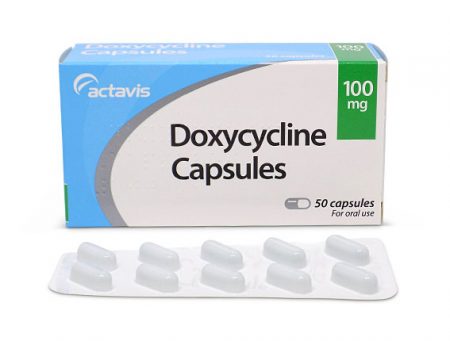
 A 2017 proof-of-concept study suggests that the antibiotic doxycycline can block the formation of negative thoughts and fear memories, perhaps offering a new way to treat or prevent post-traumatic stress disorder (PTSD).
In the study by Dominik R. Bach in the journal Molecular Psychiatry, healthy adults who received doxycycline had a lower fear response to fearful stimuli compared to healthy adults who received a placebo. The 76 participants received either doxycycline or placebo and then were taught to associate a color with an electric shock. Later, they were exposed to the color accompanied by a loud sound (but no shock), and their startle response was measured by tracking eye blinks, an instinctive response to sudden threats. Bach and colleagues found that the fear response was 60% lower in those participants who received doxycycline, suggesting that the antibiotic disrupted the fear memory linking the color to a threat.
The theory is based on evidence that doxycycline can inhibit metalloproteinase enzymes, which are involved in memory formation.
While in the study doxycycline was delivered before the fearful event occurred, there is hope that the antibiotic could also do some good after the fact. There is growing evidence that actively recalling a traumatic event can open a ‘memory reconsolidation window’ during which emotions associated with that event are open to change. Bach and colleagues hope to follow up with studies involving this reconsolidation window.
Another line of research is exploring how pain medications may reduce the emotional power of traumatic memories, because intense pain strengthens memory consolidation.
Revising Traumatic Memories in the Reconsolidation Window
 We have previously described in the BNN how therapies can take advantage of the memory reconsolidation window to reduce the power of traumatic memories. Five minutes to one hour following active emotional recall of a traumatic event, a ‘window’ opens during which therapies can revise or extinguish the traumatic memory. A 2017 article by our Editor-in-Chief Robert M. Post and Robert Kegan in the journal Psychiatric Research describes how the reconsolidation window could theoretically be used to prevent recurring depressive episodes.
The theory is based on the idea that depressive episodes initially stem from stressors, but eventually become ingrained in the brain’s habit memory system. Cognitive behavioral therapy during the memory reconsolidation window might be a good way to disrupt these habit memories.
The memory reconsolidation window has already been used successfully to reduce traumatic memories and even to reduce heroin and cocaine cravings in addiction. The idea in changing traumatic memories, in the words of researcher Göran Högberg in a 2011 article in the journal Psychology Research in Behavior Management, is to “change a reliving intruding memory into a more distant episodic memory.” Post and Kegan suggest that work in depression would have a similar goal, to rework the triggering experience and render the depressive experience “less harsh, severe, [and] self-defeating (guilt-inducing).”
In exploring this new therapeutic approach, Post and Kegan suggest that it might be best to begin with patients whose depressive episodes are triggered by stressors.
The patient would be encouraged to recall the memory of the particular stressor and any emotions related to it. Then they would be prompted to reframe the memory, either by recognizing adaptive aspects of their response, focusing on their youth at the time of the stressor in the case of childhood memories, addressing any guilt the patient may feel, or other techniques used in trauma therapy. Evoking positive feelings during this period via relaxation exercises would be another useful practice.
In addition to targeting stressors that precede depression, the stress of the depressive experience itself could be a target of reframing during the reconsolidation window.
Questions remain, such as whether to target early or more recent memories, and whether this technique would be as useful in reducing manic episodes. Patient characteristics might also affect the success of this type of therapeutic intervention.
Post and Kegan also address how the therapy might be used in different stages of illness, and how it might be combined with other therapies, such as medications or procedures such as repeated transcranial magnetic stimulation (rTMS).


