6 Minutes of Intense Cycling Produces Major Increases in BDNF
Brain derived neurotrophic factor (BDNF) is necessary for new synapses and call survival. A new study in J. Physiology (2023) reports that the increases in BDNF from short intense cycling exercise are much greater than from prolonged (90-minute) light cycling. The authors think that this is cause by the increases in lactate produced which helps up regulate BDNF production. This could be good for fighting depression and Alzheimer’s disease, where BDNF levels are low.
Bottom line: If you don’t have much time, bust your buns.
Teens with Bipolar Disorder at Increased Risk for Cardiovascular Disease
 A scientific statement from the American Heart Association reported in 2015 that youth with major depressive disorder and bipolar disorder are at moderate (Tier II level) increased risk for cardiovascular disorders. The combined prevalence of these illnesses in adolescents in the US is approximately 10%.
There are many factors that contribute to this risk, including inflammation, oxidative stress (when the body falls behind neutralizing harmful substances produced during metabolism), dysfunction in the autonomic nerve system, and problems with the endothelium (the inner lining of blood vessels). Lifestyle factors include adversity in early life, sleep disturbance, sedentary lifestyle, poor nutrition, and abuse of tobacco, alcohol, or other substances.
Taking some atypical antipsychotics as treatment for bipolar disorder also contributes to the risk of cardiovascular problems by increasing weight and/or lipid levels. Among the atypicals, ziprasidone (Geodon) and lurasidone (Latuda) come with the lowest likelihood of weight gain.
The statement by Benjamin I. Goldstein and colleagues that appeared in the Heart Association-affiliated journal Circulation suggested that therapeutic interventions should address some of these risk factors to help prevent cardiovascular problems and improve life expectancy for young people with depression or bipolar disorder. These could include a good diet, regular exercise, and treatments with good long-term tolerability that are aimed at preventing episodes.
The Role of Inflammatory Markers and BDNF
Inflammation worsens the risk of cardiovascular problems, while brain-derived neurotrophic factor (BDNF), which protects neurons and plays a role in learning and memory, may improve prospects for someone with depression or bipolar disorder.
A 2017 article by Jessica K. Hatch and colleagues including Goldstein in the Journal of Clinical Psychiatry suggests that inflammation and BDNF are mediators of cardiovascular risk in youth with bipolar disorder. The study looked at 40 adolescents with bipolar disorder and 20 healthy controls.
Those with bipolar disorder had greater waist circumference, body mass index, and pulse pressure than the controls. The youth with bipolar disorder also had higher levels of the inflammatory cytokine Il-6. Participants who had lower BDNF had greater thickness of the carotid vessel internal lining (intima media).
Hatch and colleagues point to the importance of prevention strategies in adolescents with these indicators of increased cardiovascular risk. These data complement the American Heart Association’s recognition of adolescent mood disorders as a large problem that deserves wider attention both in psychiatry and in the media.
Early Cannabis Use and BDNF Gene Variant Increase Psychosis Risk
 Normal variations in genes can affect risk of mental illness. One gene that has been implicated in psychosis risk is known as BDNF. It controls production of brain-derived neurotrophic factor, a protein that protects neurons and is important for learning and memory. Another important gene is COMT, which controls production of the enzyme catechol-O-methyltransferase, which breaks down neurotransmitters such as dopamine in the brain.
Several forms of these genes appear in the population. These normal variations in genes are known as polymorphisms. Certain polymorphisms have been linked to disease risk. A study by Anna Mané and colleagues published in the Journal of Psychiatric Research in 2017 explored links between COMT and BDNF polymorphisms, cannabis use, and age at first episode of psychosis.
Mané and colleagues found that among 260 Caucasians being treated for a first episode of psychosis, the presence of a BDNF polymorphism known as val-66-met and a history of early cannabis use were associated with younger age at psychosis onset.
The val-66-met version of BDNF occurs in 25-35% of the population. It functions less efficiently than a version called val-66-val.
The researchers also found that males were more likely to have used cannabis at a young age.
Editor’s Note: In the general population, marijuana use doubles the risk of developing psychosis. Previous data had indicated that the risk was higher for those with a COMT polymorphism known as val-158-val that leads to more efficient metabolism of dopamine in the prefrontal cortex. The resulting deficits in dopamine increase vulnerability to psychosis compared to people with the val-158-met version of the COMT gene.
The new study by Mané and colleagues suggests that a common form of BDNF may be associated with an earlier onset of psychosis. Bottom line: Pot is dangerous for young users and can induce psychosis, particularly in people at genetic risk. Pot may be legal in many places, but heavy use in young people remains risky for mental health and cognitive functioning.
The company Genomind offers genetic testing for BDNF and COMT variants as part of a routine panel.
Low Vitamin D Linked to Small Hippocampus & Schizophrenia
Low levels of vitamin D have been linked to schizophrenia in several studies. In one, infants with low vitamin D were more likely to develop schizophrenia in adulthood, but supplementation reduced this risk. A 2015 article by Venkataram Shivakumar and colleagues in the journal Psychiatry Research: Neuroimaging found that among patients with schizophrenia who were not currently taking (or in some cases, had never taken) antipsychotic medication, low levels of vitamin D were linked to smaller gray matter volume in the right hippocampus, an area involved in schizophrenia.
Vitamin D has neuroprotective effects and is important to normal brain development and function. Vitamin D is essential to the production of brain-derived neurotrophic factor (BDNF), a protein that is important for learning and memory, and vitamin D also reduces oxidative stress. BDNF deficiency and oxidative stress have both been linked to schizophrenia, and they both can cause abnormalities in the hippocampus.
Antidepressant Effects of Sleep Deprivation are Associated with Increases in BDNF
 Brain-derived neurotrophic factor (BDNF) is a protein that protects neurons and aids with learning and memory. BDNF levels are low in people with depression, and every type of antidepressant treatment increases BDNF, including different types of medications, electro-convulsive therapy, repeated transcranial magnetic stimulation (rTMS), exercise, atypical antipsychotic drugs used to treat bipolar depression, omega-3 fatty acids, and ketamine. New research shows that sleep deprivation, which is sometimes used to rapidly improve depression, also increases BDNF.
A study by researcher Anne Eckert and colleagues found that partial sleep deprivation (preventing sleep only during the second half of one night) increased BDNF levels in the blood the following day. Those participants who responded well to the sleep deprivation were found to have BDNF levels that fluctuated more throughout the day before the sleep deprivation compared to participants who did not respond well to the sleep deprivation, whose BDNF levels were relatively stable.
The research, which was presented at a scientific meeting in 2015, suggests that sleep deprivation in depressed patients increases BDNF, and that such increases are an important function of any antidepressant treatment.
Reduced Cognitive Function and Other Abnormalities in Pediatric Bipolar Disorder
 At the 2015 meeting of the International Society for Bipolar Disorders, Ben Goldstein described a study of cognitive dysfunction in pediatric bipolar disorder. Children with bipolar disorder were three years behind in executive functioning (which covers abilities such as planning and problem-solving) and verbal memory.
There were other abnormalities. Youth with bipolar disorder had smaller amygdalas, and those with larger amygdalas recovered better. Perception of facial emotion was another area of weakness for children (and adults) with bipolar disorder. Studies show increased activity of the amygdala during facial emotion recognition tasks.
Goldstein reported that nine studies show that youth with bipolar disorder have reduced white matter integrity. This has also been observed in their relatives without bipolar disorder, suggesting that it is a sign of vulnerability to bipolar illness. This could identify children who could benefit from preemptive treatment because they are at high risk for developing bipolar disorder due to a family history of the illness.
There are some indications of increased inflammation in pediatric bipolar disorder. CRP, a protein that is a marker of inflammation, is elevated to a level equivalent to those in kids with juvenile rheumatoid arthritis before treatment (about 3 mg/L). CRP levels may be able to predict onset of depression or mania in those with minor symptoms, and is also associated with depression duration and severity. Goldstein reported that TNF-alpha, another inflammatory marker, may be elevated in children with psychosis.
Goldstein noted a study by Ghanshyam Pandey that showed that improvement in pediatric bipolar disorder was related to increases in BDNF, a protein that protects neurons. Cognitive flexibility interacted with CRP and BDNF—those with low BDNF had more cognitive impairment as their CRP increased than did those with high BDNF.
Genetic Variation Predicts Which Type of Antidepressant Will Be Effective
 In a six-month study of Caucasian patients, normal variations in the gene that is responsible for brain-derived neurotrophic factor (BDNF) predicted whether patients would respond better to a selective serotonin reuptake inhibitor (SSRI) antidepressant versus a serotonin and norepinephrine reuptake inhibitor (SNRI) or a tricycle antidepressant. There are several common variants of the BDNF gene, depending on which types of amino acids appear in its coding—valine or methionine. Patients with the most common version, two valines (or Val66Val), responded better to SSRIs. About two-thirds of the population has this version of the gene, which functions most efficiently. The remaining third have at least one methionine in the BDNF gene. Patients with a Met variation responded better to SNRIs and tricyclic antidepressants.
The study by R. Colle and colleagues was published in the Journal of Affective Disorders in 2015. Of the patients who were prescribed SSRIs, 68.1% of patients with the Val/Val version responded to the medication after three months, compared to 44% of the patients with a Met version. Of patients prescribed SNRIs or tricyclics, 60.9% of the Met patients reached remission by six months, compared to only 33.3% of the Val/Val patients.
Editor’s Note: In an earlier BNN we reported that according to research published by Gonzalo Laje and colleagues in the journal Biological Psychiatry in 2012, depressed patients with the better functioning Val66Val allele of BDNF respond best to ketamine, while those with the intermediate functioning Val66Met allele respond less well.
Lithium Extends Anti-Depressant Effects of Ketamine in Mice
While it can sometimes take weeks for the effects of antidepressant treatments to appear, intravenous ketamine can produce antidepressant effects in as little as two hours. However, ketamine’s effects fade after three to five days. New animal research by Chi-Tso Chiu et al. explores whether adding lithium to ketamine treatment can produce more sustained antidepressant effects.
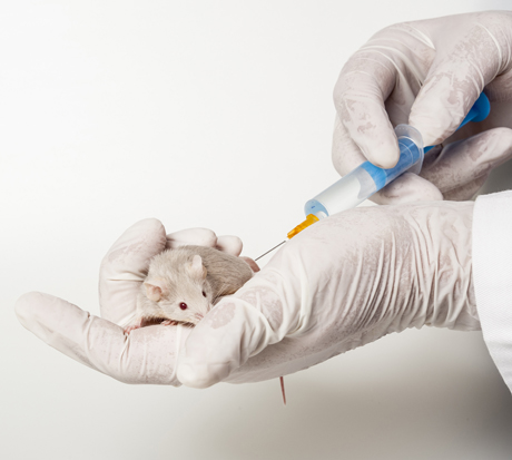
Mice who are restrained by being placed in a tube for several hours (chronic restraint stress) exhibit a behavioral and neurochemical profile that resembles human depression. When Chiu and colleagues pretreated these stressed mice with sub-therapeutic doses of lithium (600 mg/L) in their drinking water for several weeks, a sub-therapeutic dose of ketamine (2.5 mg/kg of body weight) was enough to produce robust antidepressant effects in the mice, while neither drug alone was effective at these doses.
The combination of ketamine and lithium also restored the density of spines on the dendrites of neurons in the medial prefrontal cortex. Post-treatment with lithium (1200 mg/L) for several weeks was also successful in extending the effects of a single (50 mg/kg) ketamine injection.
Both lithium and ketamine affect the intracellular signaling pathway mTOR. Ketamine activates the pathway, increasing levels of synaptic proteins and dendritic spine density. It also increases brain-derived neurotrophic factor (BDNF) and the BDNF receptor TrkB. BDNF is important for learning and memory.
When lithium was added to the treatment of the mice with ketamine, the mTOR and BNDF pathways were further activated. Lithium also inhibits the receptor GSK-3, supporting ketamine’s rapid-acting antidepressant effects.
Ketamine treatment can produce oxidative stress, in which toxic free radicals can endanger cells, and the addition of low doses of lithium also completely prevented this neurochemical side effect.
Chiu and colleagues hope that the findings of this study in mice can eventually be applied to research in humans in the hopes of finding a clinical option that would sustain the rapid-onset antidepressant effects of ketamine for the long term.
Exercise Helps Mice with Spacial Learning
Exercise increases brain-derived neurotrophic factor (BDNF), a protein that protects neurons and is important for learning and memory. In a study of mice who were trained to find objects, sedentary mice could not discriminate between familiar object locations and novel ones 24 hours after receiving weak training, while mice who had voluntarily taken part in exercise over a 3-week period could easily distinguish between these locations after the weak training.
Mice who received sodium butyrate (NaB) after training behaved similarly well to those who had exercised. Sodium butyrate is a histone deacetylase (HDAC) inhibitor, meaning it helps keep acetyl groups on histones, around which DNA is wrapped, making the DNA easier to transcribe. In this case the easy transcription of DNA enables learning under conditions in which it might not usually take place.
Both sodium butyrate and exercise promote learning through their effects on BDNF in the hippocampus. They make the DNA for BDNF easier to transcribe, suggesting that exercise can put the brain in a state of readiness to create new or more lasting memories.
How Inflammation Increases Glutamate Overexcitation And Neurotoxicity
Research has shown a link between inflammation and mental illness. Inflammation leads to a series of chemical changes that can overexcite neurons and interfere with the protection of neurons.
Inflammation increases the production of indoleamine-pyrrole 2,3-dioxygenase (IDO), an enzyme that breaks down the amino acid tryptophan into kynurenic acid and quinolinic acid. They in turn increase glutamate, the main excitatory neurotransmitter, and decrease brain-derived neurotrophic factor (BDNF), which keeps neurons healthy.
Kynurenic acid stimulates microglia, which clean up the central nervous system as a form of immune defense, to produce inflammatory cytokine proteins.
Quinolinic acid directly stimulates glutamate receptors and encourages glutamate release from astrocytes. Quinolinic acid also blocks glutamate removal that would normally occur through reuptake into the astrocytes, leading to more stimulation of extrasynaptic glutamate receptors and decreases in BDNF.
Quinolinic acid’s effects are opposite to those of the antidepressant ketamine, which blocks glutamate NMDA receptors and increases BDNF. When people are given interferon protein for the treatment of cancers, quinolinic acid increases in cerebrospinal fluid, inducing depression. The severity of depression induced is correlated with the patient’s levels of quinolinic acid.
It appears that ketamine has indirect anti-inflammatory effects through its ability to block glutamate receptors and increase BDNF.


