Cinnamon Improves Memory in Mice
A recent study found that mice that ate more cinnamon got better and faster at learning. In the study, published in the Journal of Neuroimmune Pharmacology in 2016, separated mice into good learners and poor learners based on how easily they navigated a maze to find food. After the poor learners were fed cinnamon for a month, they could find the food more than twice as quickly as before.
The benefits of cinnamon come from sodium benzoate, a chemical produced as the body breaks down the cinnamon. Sodium benzoate enters the brain and allows the hippocampus to create new neurons.
Feeding cinnamon to the poor learning mice normalized their levels of receptors for the neurotransmitter GABA, closing the gap with good learners. Sodium benzoate also improved the structural integrity of some brain cells. Cinnamon also can help sensitize insulin receptors.
Doctors hope these findings may eventually contribute to treatment research on Alzheimer’s and Parkinson’s diseases.
Cinnamon should be consumed in moderate quantities because the Chinese variety most commonly found in North American supermarkets has high levels of coumarin, a compound that can be toxic to the liver when consumed in large quantities. Ceylon (Sri Lankan) cinnamon has lower levels of coumarin.
Guanfacine Improves ADHD Symptoms and Academic and Social Functioning in Children
A study by researcher J.H. Newcorn and colleagues published in the Journal of the American Academy of Child and Adolescent Psychiatry in 2013 found that eight weeks of treatment with the drug guanfacine (extended release) improved symptoms of attention deficit hyperactivity disorder (ADHD) in North American children compared to placebo. A 2015 study by M.A. Stein and colleagues in the journal CNS Drugs extended this research, determining that guanfacine also improved academic and social functioning, including family dynamics, in the same group of children.
Children aged 6–12 who had been diagnosed with ADHD received either placebo or 1 to 4 mg of guanfacine extended release either in the morning or evening. The children in both guanfacine groups showed improvements in family interactions, learning and school, social behavior, and risky behavior compared to those taking placebo. No improvements were seen in life skills or self-concept. The improvements in functioning were linked to the drug’s effectiveness in improving ADHD symptoms. Those children whose ADHD symptoms improved on guanfacine were also more likely to see improvements in academic and social functioning.
Exercise Helps Mice with Spacial Learning
Exercise increases brain-derived neurotrophic factor (BDNF), a protein that protects neurons and is important for learning and memory. In a study of mice who were trained to find objects, sedentary mice could not discriminate between familiar object locations and novel ones 24 hours after receiving weak training, while mice who had voluntarily taken part in exercise over a 3-week period could easily distinguish between these locations after the weak training.
Mice who received sodium butyrate (NaB) after training behaved similarly well to those who had exercised. Sodium butyrate is a histone deacetylase (HDAC) inhibitor, meaning it helps keep acetyl groups on histones, around which DNA is wrapped, making the DNA easier to transcribe. In this case the easy transcription of DNA enables learning under conditions in which it might not usually take place.
Both sodium butyrate and exercise promote learning through their effects on BDNF in the hippocampus. They make the DNA for BDNF easier to transcribe, suggesting that exercise can put the brain in a state of readiness to create new or more lasting memories.
BDNF in Learning and Memory
Brain-derived neurotrophic factor (BDNF) is involved in various aspects of learning and memory. The DNA for BDNF contains nine different regulatory sites, each of which is involved in different aspects of learning. Researcher Keri Martinovich studied each site by selectively knocking each one out with a genetic manipulation. She found that blocking the e1 site increased acquisition of new learning and recall in mice, while e2 did the opposite. Blockade of e4 had no effect on these memory functions but markedly blocked the process of extinction, which involves a different kind of new learning.
A mouse that learned to associate a particular cue with a shock (a process known as conditioned fear) will stop reacting to the cue after it is presented many times without a shock. This learning that the cue is no longer associated with the shock is referred to as extinction. The animals with e4 blocked in their BDNF did not develop the new extinction learning, and continued to react to the cue as if it were still associated with the shock.
Editor’s Note: These data may have clinical relevance for humans. The anticonvulsant valproate (trade name Depakote), a histone deacetylase inhibitor, selectively increases the e4 promoter site of BDNF and facilitates extinction of conditioned fear, according to research by Tim Bredy et al. published in 2010.
Clinical trails should examine whether valproate could enhance fear extinction in patients with post-traumatic stress disorder (PTSD).
Rats Learn Fear Conditioning From One Another
Rats who are taught to associate a light with an electric shock learn to avoid the light. This process is known as conditioned fear. New research shows that if one rat watches another rat go through fear conditioning, the observing rat will also show the effects of fear conditioning. It will also avoid the light, but only if it had previous experience with fear conditioning. It appears that rats have the ability to learn from other rats’ painful experiences.
Psychiatric Revolution: Changes in Behavior Are Associated with Dendritic Spine Shape and Number
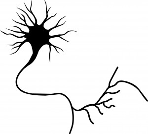
New research shows that cocaine, defeat stress, the rapid-acting antidepressant ketamine, and learning and memory can change the size, shape, or number of spines on the dendrites of neurons. Dendrites conduct electrical impulses into the cell body from neighboring neurons.
Cocaine
Several researchers, including Peter Kalivas at the Medical University of South Carolina, have reported that cocaine increases the size of the spines on the dendrites of a certain kind of neurons (GABAergic medium spiny neurons) in the nucleus accumbens (the reward center in the brain). This occurs through a dopamine D1 selective mechanism. N-acetylcysteine, a drug that can be found in health food stores, decreases cocaine intake in animals and humans, and also normalizes the size of dendritic spines.
Depression
Depression in animals and humans is associated with decreases in Rac1, a protein in the dendritic spines on GABA neurons in the nucleus accumbens. Rac1 regulates actin and other molecules that alter the shape of the spines.
In an animal model of depression called defeat stress, rodents are stressed by repeatedly being placed in a larger animal’s territory. Their subsequent behavior mimics clinical depression. This kind of social defeat stress decreases Rac1 and causes spines to become thin and lose some function. Replacing Rac1 returns the spines to a more mature mushroom shape and reverses the depressive behavior of these socially defeated animals. Researcher Scott Russo has also found Rac1 deficits in the nucleus accumbens of depressed patients who committed suicide. Russo suggests that decreases in Rac1 are responsible for the manifestation of social avoidance and other depressive behaviors in the defeat stress animal model, and that finding ways to increase Rac1 in humans would be an important new target for antidepressant drug development.
Another animal model of depression called chronic intermittent stress (in which the animals are exposed to a series of unexpected stressors like sounds or mild shocks) also induces depression-like behavior and makes the dendritic spines thin and stubby. The drug ketamine, which can bring about antidepressant effects in humans in as short a time as 2 hours, rapidly reverses the depressive behavior in animals and converts the spines back to the larger, more mature mushroom-shape they typically have.
Learning and Extinction of Fear
Researcher Wenbiao Gan has reported that fear conditioning can change the number of dendritic spines. When animals hear a tone paired with an electrical shock, they begin to exhibit a fear response to the tone. In layer 5 of the prefrontal cortex, spines are eliminated when conditioned fear develops, and are reformed (near where the eliminated spines were) during extinction training, when animals hear the tones without receiving the shock and learn not to fear the tone. However, in the primary auditory cortex the changes are opposite: new spines are formed with learning, and spines are eliminated with extinction.
Editor’s Note: It appears that we have arrived at a new milestone in psychiatry. In the field of neurology, changes seen in the brains of patients with strokes or Alzheimer’s dementia have been considered “real” because cells were obviously lost or dead. Psychiatry, in comparison, has been considered a soft science because neuronal changes have been more difficult to see and illnesses were and still are called “mental.” Now that new technologies have made a deeper level of precision, observation, and analysis possible, we know that the brain’s 12 billion neurons and 4 times as many glial cells are exquisitely plastic–capable of biochemical and structural changes that can be reversed using appropriate therapeutic maneuvers.
The changes associated with abnormal behaviors, addictions, and even normal processes of learning and memory now have clearly been shown to correspond with the size, shape, and biochemistry of dendritic spines. These subtle, reproducible changes in the brain and body are amenable to therapeutic intervention, and are now even more demanding of sophisticated medical attention.
Opening The Reconsolidation Window to Extinguish Fear Memories: A New Conceptual Approach to Psychotherapeutics
 Memory processes occur in several phases. Short-term memory is converted to long-term memory by a process of consolidation that requires the synthesis of new proteins. Transcription factors in the nuclei of hundreds of millions of nerve cells are activated so that specific synapses can be modified for the long term. If protein synthesis is inhibited during a period within a few hours after new learning has occurred, what was learned never gets consolidated and is essentially forgotten. It is thought that this phase of consolidation happens when a memory trace moves from short-term storage in the hippocampus to long-term storage in the cerebral cortex.
Recently a later phase of memory storage called reconsolidation has been identified. When an old memory is recalled, the reconsolidation window opens, and the memory trace becomes temporarily amenable to change. The reconsolidation window (the period during which the trace can be revised) is thought to begin five minutes after a memory is recalled and last for an hour or possibly two. New learning that takes place during the reconsolidation window can be more profound than learning that occurs without recall of the related memory or after the reconsolidation window has closed.
Consider the example of a fearful memory created when a person is attacked in a dark alley. If the person repeatedly visits the same alley without being attacked, they can eventually become less afraid of dark places. Repeated viewing of pictures of dark places can also extinguish the fear. These are typical ways in which a fear memory is extinguished. However, the original fear is subject to spontaneous recovery (the fear of dark places returns without provocation) or to reactivation (if another dangerous situation is encountered, the person may regain their fear of dark alleys).
The new findings suggest that if the extinction process (the repeated exposures to the pictures of the dark alley) takes place during the reconsolidation window after the fear memory of being attacked is recalled, the old fear can be permanently reversed (wiped clean, or re-edited such that it appears forgotten) so that it is no longer subject to spontaneous recovery or reactivation.
Editor’s Note: To accomplish extinction training within the reconsolidation window, first a person must actively recall the old memory, opening the reconsolidation window. Then, after a 5-minute delay, extinction training (e.g. new learning that the old feared place is now safe) should take place within the next hour. This process has been demonstrated in animal studies and is thought to be clinically relevant for humans in the case of phobic anxiety and post-traumatic stress disorder (PTSD). The psychotherapeutic implications of using the reconsolidation window to better ameliorate PTSD fears, avoidance, and flashbacks are enormous.
Observing the Amygdala’s Role in the Extinction of Fear Memory Traces
The amygdala is a crucial part of the learned or conditioned fear pathway. It is activated during fear conditioning and during the recall of cues associated with the fear experience. If the amygdala is removed, conditioning fear does not occur.
A new study published in Science this year by Agren et al. indicates that in humans, the amygdala-based response to conditioned fear can be completely abolished using extinction training within the memory reconsolidation window. Training that took place 10 minutes after the fear memory was activated was successful, while training that took place 6 hours later was not. Read more
Exercise Good for Learning and Memory in Children and the Elderly
Another article in the Telegraph today suggests that aerobic exercise can increase the size of the hippocampus in elderly people and lead to improvements in memory, attention, and ability to multi-task. Children who were more fit were also better at multitasking. Art Kramer of the Beckman Institute for Advanced Science and Technology at the University of Illinois said,
“It is aerobic exercise that is important so by starting off doing just 15 minutes a day and working up to 45 minutes to an hour of continuous working we can see some real improvements in cognition after six months to a year.
“We have been able to do a lot of neuroimaging work alongside our studies in the elderly and show that brain networks and structures also change with exercise.


