
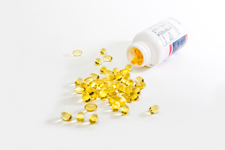
Omega-3 Fatty Acids Improve Mood and Limbic Hyperactivity in Youth with Bipolar Depression
Children who have a parent with bipolar disorder are at risk for bipolar illness, but it may first present as depression. Treating these children with antidepressants has the risk of bringing on manic episodes. Researchers are looking for treatment options for youth at risk for bipolar disorder.
Robert McNamara and colleagues found that 12 weeks of omega-3 fatty acids (2,100 mg/day) significantly improved response rates in medication-free youth ages 9–20 years compared to placebo (64% versus 36%). Omega-3 fatty acids but not placebo also reduced the activation of limbic structures in the brain (the left parahippocampal gyrus) in response to emotional stimuli.
Editor’s Note: These data add to the literature on the positive effects of 1–2 grams of omega-3 fatty acids in depression. Given the safety of omega-3 fatty acids and the ambiguous effects of antidepressants in bipolar depression, omega-3 fatty acids would appear to a good alternative, especially since the FDA-approved atypical antipsychotics (quetiapine and lurasidone) are not approved for bipolar depression in people under age 18.
Cariprazine More Potent Than Aripiprazole at Dopamine D3 Receptors
Cariprazine is a new atypical antipsychotic that has positive results in the treatment of schizophrenia, bipolar mania and depression, and as an add-on treatment to antidepressants in unipolar depression. In a recent study by Frank Tarazi and colleagues, cariprazine shared some actions with aripiprazole (trade name Abilify), which is approved by the Federal Drug Administration as an adjunctive treatment for unipolar depression in addition to treating mania in bipolar disorder and schizophrenia.
Both cariprazine and aripiprazole partially block dopamine receptors. They allow a little stimulation of the receptors (activating or agonist effects) while preventing dopamine from binding there. With ongoing treatment, this blocking action prompts an increase in the number of dopamine receptors to compensate for the blocking.
In rats, both drugs led to increases in several types of dopamine receptors, but cariprazine did so at lower doses. Following 28 days of treatment with aripiprazole at doses of 5–15 mg/kg, rats had higher levels of D2 and D4 receptors in several brain regions than other rats that received no drug. Higher doses of aripiprazole also increased D3 receptors in some regions, indicating that the drug works less well at those receptors. Lower doses of cariprazine (0.06–0.6 mg/kg) increased D2 and D4 receptors significantly, and increased D3 receptors more than aripiprazole did, and in a greater number of forebrain regions.
Among antipsychotic drugs, both aripiprazole and cariprazine are unique in providing a little stimulation (partial agonist effects) at dopamine receptors, while all others drugs in the class are pure blockers (antagonists) of dopamine receptors.
The researchers concluded that cariprazine was more potent than aripiprazole at dopamine receptors, especially D3 receptors.
Brain Activity Differentiates Youth with Bipolar Disorder from Youth with Unipolar Depression

Both bipolar disorder and unipolar depression often begin in childhood or adolescence, but it can be difficult to distinguish the two using symptoms only. People with bipolar illness may go a decade without receiving a correct diagnosis. Researcher Jorge Almeida and colleagues recently performed a meta-analysis of previous studies to determine what neural activity is typical of children with bipolar disorder versus children with unipolar depression while processing images of facial emotion. They found that youth with bipolar disorder were more likely to show limbic hyperactivity and cortical hypoactivity during emotional face processing than youth with unipolar depression. Almeida and colleagues hope that this type of data may eventually be used to diagnose these disorders or to measure whether treatment has been successful.
Antipsychotics That Worked for A First Episode May Not Work As Well a Second Time
 In new research by Ofer Agid and colleagues, patients in their first schizophrenic episode who reached remission in response to one of two antipsychotic medications (risperidone or olanzapine) and relapsed due to medication non-adherence were re-treated with the same medication regimen that had brought about remission. Reinitiating the same treatment was not as successful in bringing about remission of the patients’ second psychotic episodes.
Patients showed different types of trajectories in their first remission, from immediate to gradual improvement, and these predicted parallel trajectories of their treatment response during the second episode, though the muted response to antipsychotics existed across the board. Dopamine is the main target of antipsychotic treatments, but its role in schizophrenia is not straightforward, and Agid and colleagues stress that response and relapse are multidimensional processes.
Editor’s Note: These data are consistent with the research of J.A. Lieberman and colleagues fifteen years ago, which showed that response to antipsychotic treatment is poorer in successive episodes of psychosis. The findings are also consistent with the idea of episode sensitization in mood disorders, developed by this author (Robert Post). Episode sensitization refers to the case in which greater numbers of prior depressions or manias are associated with faster relapse and a greater degree of treatment resistance.
The data raise major doubts about the common practice of quitting medications to see if remission can be maintained without them. There are dozens of studies in patients with schizophrenia showing that continuous treatment is more effective than intermittent treatment.
White Blood Cells Can Convey Resilience to Stress
white blood cells
Mice subjected to chronic defeat stress (being placed in the home cage of a larger, more aggressive mouse) behave in ways that resemble human anxiety and depression. In new research by Miles Herkenham and colleagues at the National Institute of Mental Health, in which they explored the adaptive immune system’s affect on mood, mice exposed to this type of stress showed increases in inflammatory cytokines in the blood (including TNFalpha, IL-1beta, IL-2, IL-3, IL-6, IL-17, and IFNgamma) compared to a control group. Interestingly, when white blood cells (lymphocytes) from stressed animals were transferred to a new set of animals, the recipient mice seemed to benefit from greater resilience to stress in a variety of ways.
Mice that received white blood cells from defeat-stressed animals had lower levels of TNFalpha, IL-1beta, IL-2, IL-3, and IL-17 than a control group that received white blood cells from unstressed mice. The recipient mice also exhibited reduced anxious and depressive behaviors in a litany of behavioral tests compared to both the group that received white blood cells from unstressed mice and a group that received a saline injection instead. Lastly, the recipients of the white blood cells from stressed animals showed more new neurons in the dentate gyrus of the hippocampus. (Hippocampal neurogenesis is decreased by stressors and increased by antidepressants.)
Herkenham and colleagues concluded that psychopathology is not just a downward spiral—the immune system plays an active role in adapting to stress, with lymphocytes being programmed by stress to provide antidepressant functions.
Editor’s Note: These data add a new twist to the studies of Scott Russo, who found that IL-6 secreted by white cells of animals subject to defeat stress was the cause of the depressive-like behaviors they exhibited. If IL-6 was blocked, the behaviors did not occur. Now it would appear from Herkenham’s work that something about the timing, the type of cytokines, or the transfer of the white cells conveyed protective antidepressant-like effects in this case.
The Combination of N-Acetylcysteine and Varenicline Reduces Nicotine Addiction in Rats
Nicotine addiction is highly cue-dependent, meaning that certain situations or places will make smokers crave a cigarette even if they’re trying to quit. Researchers working with rodents are exploring a combination of treatments that address different behavioral and neurobiological mechanisms to reduce nicotine addiction. In a recent study by Cassandra Gipson-Reichardt and colleagues, N-acetylcysteine reduced cue-induced nicotine seeking, while varenicline reduced nicotine self-administration. Together the drugs worked better to reduce nicotine relapse than either drug on its own.
In the study, rats were trained to self-administer nicotine (with 0.02mg/kg infusions), and cues were used to reinstate nicotine seeking. The rats were treated with 10 and 30 mg/kg injections of NAC and 1 and 3 mg/kg injections of varenicline.
Relapse is associated with rapid synaptic potentiation in the reward area of the brain, the nucleus accumbens. In addition to the positive behavioral changes noted, NAC also inhibited this synaptic potentiation, limiting rapid changes in the size of spines on dendrites and reducing the ratio of AMPA to NMDA (two different compounds that mimic glutamate) in the core of the nucleus accumbens.
Editor’s Note: The combination of NAC and varenicline has not yet been studied in humans, but because both compounds are effective in reducing smoking, it is likely that this animal research on nicotine will be replicated in humans who are addicted to the nicotine in cigarettes.
N-Acetylsysteine Reduces Cutting Behaviors

Cutting, or non-suicidal self injury, is a serious problem among adolescents, and few treatments are available. Researcher Kathryn Cullen and colleagues have found that N-acetylcysteine (NAC), an antioxidant nutritional supplement that has been effective in the treatment of depression and many addictions and habit-related behaviors, can reduce cutting.
The study included 25 participants with a history of non-suicidal self injury, aged 13–21, and 12 controls. They participated in brain scans before and after treatment. Compared to the controls, the self-injurers showed greater overall psychopathology, greater activation in a few brain regions (precuneus, posterior cingulate, insula, and temporal lobes), and reduced lower left frontal activation. Patients who received NAC up to 900mg twice daily in weeks 5–8 of the study reduced their cutting and also showed reduced psychopathology. An increase in frontal activation in response to negative emotion was linked to the reduction in cutting.
Editor’s Note: NAC improves mood in depression, many addictions, and many habits including trichotillomania (excessive hair-pulling), nail biting, and cutting. It may do this by increasing glial glutamate transporters in the nucleus accumbens, the brain’s reward center, which lessens the magnitude of the glutamate signal, mediating the compulsion to engage in the habitual behavior.
Connections Between Stress and Substance Use May Be Mediated By Corticotropin-Releasing Factor
 Researchers hope to map out the neurocircuitry by which stress leads to compulsive drug taking. A recent study by Klaus Miczek and colleagues examined different rodents’ responses to the stress of being repeatedly placed in the cage of a larger, more aggressive rodent, developing what is known as defeat stress, a set of behaviors that mimic human depression. Mice and rats showed increases in the stress hormone corticosterone that did not diminish over repeated run-ins with a larger animal. Rodents who were exposed to this stress became sensitized to cocaine or amphetamine, showing hyperactivity that increased each time they accessed the drug (the opposite of a tolerance response). Some also “binged” on cocaine, which they were able to self-administrate by pushing a lever to receive infusions. The mice and rats that went through the social defeat showed elevated levels of dopamine in the nucleus accumbens, the brain’s reward center. Levels were related to the severity of their stressful experience.
Later the rodents had a choice between water and a 20% alcohol solution. The researchers determined what type of stress led the rodents to consume the alcohol solution instead of the water. The maximal effect was seen in two types of mice that suffered an attack of less than five minutes that resulted in a moderate number of attack bites (30); this resulted in the mice consuming large amounts (15–30 g/kg/day) of the alcohol solution. Earlier sensitization to cocaine or amphetamine did not predict later alcohol or cocaine self-administration.
When the researchers injected the rodents with antagonists of the receptors for corticotropin-releasing factor, a hormone and neurotransmitter important in stress response, prior to each episode of social defeat, the rodents did not escalate their cocaine or alcohol self-administration, indicating that CRF plays an essential role in the process by which stress makes animals prone to using substances.
In related research by Camilla Karlsson and colleagues, IL-1R1 and TNF-1R, the receptors for two inflammatory cytokines, mediated the effects of social stress on escalated alcohol use in mice.
Childhood Adversity, Gene Methylation, and Internalizing Behaviors Linked

Life experiences such as adversity in childhood have been linked to epigenetic changes to DNA. These changes do not affect the sequence of DNA, but can change how tightly DNA is wound, and thus how easily it is transcribed. One epigenetic change that can occur following adversity in childhood is methylation of the gene for the glucocorticoid receptor (NR3C1). A recent study by Kathryn Ridout and colleagues examined links between early adversity, methylation of this gene, and behavioral problems in childhood. Adversity was linked to methylation of the gene at exons 1D and 1F in the promoter of NR3C1. Methylation of the gene was associated with internalizing behaviors (e.g. depression, anxiety) but not externalizing behaviors (e.g. attention deficit hyperactivity disorder (ADHD) or oppositional defiant disorder) in children of preschool age. The NR3C1 methylation was a significant mediator of the internalizing behaviors in children who had experienced adversity.
Editor’s Note: Similar associations of methylation of the glucocorticoid receptor with childhood adversity have been reported in other clinical and animal studies and provide a mechanism for the long-lasting adverse effects of stressors in childhood.
Output From the Amygdala Mediates Reward or Fear Memories
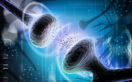
synapse
People and animals can rapidly learn to associate environmental stimuli with positive or negative outcomes, learning what to approach or avoid as they go through daily life. The amygdala plays a role in this type of emotional learning, which can be disrupted by mood disorders. In new research, Praneeth Namburi and colleagues determined that activity at the synapses in the basolateral amygdala reveals differences in the creation of fear memories and reward memories.
In animals trained with reward and fear conditioning tasks, photostimulation of neurons that then travel from the basolateral amygdala complex to the nucleus accumbens (the brain’s reward center) is positively reinforcing, while photostimulation of neurons that will travel from the basolateral amygdala complex to the centromedial nucleus of the amygdala causes aversion. There are genetic differences between the two types of neurons, including a difference in the gene for the neurotensin-1 receptor. The researchers found that neurotensin, a neuropeptide, modulates glutamate’s effect on neurons, causing some to project to the nucleus accumbens and some to project to the centromedial nucleus of the amygdala.
The researchers wrote that the results “provide a mechanistic explanation, on both a synaptic and circuit level, for how positive and negative associations can be rapidly formed, represented, and expressed within the amygdala.”
Editor’s Note: The amygdala’s creation of opposing outputs may provide clues to the mechanisms behind mania (involving the nucleus accumbens) and depression (involving the centromedial nucleus of the amygdala).
