Over-Pruning of Synapses May Explain Schizophrenia
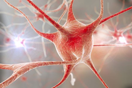
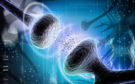
A gene that plays a role in the pruning of synapses has been linked to schizophrenia. The gene encodes an immune protein called complement component 4 (C4), which may mediate the pruning of synapses, the connections between neurons. Researchers led by Aswin Sekar found that in mice, C4 was responsible for the elimination of synapses. The team linked gene variants that lead to more production of C4A proteins to excessive pruning of synapses during adolescence, the period during which schizophrenia symptoms typically appear. This may explain why the brains of people with schizophrenia have fewer neural connections. The researchers hope that future therapies may target the genetic roots of the illness rather than simply treating its symptoms.
Output From the Amygdala Mediates Reward or Fear Memories
People and animals can rapidly learn to associate environmental stimuli with positive or negative outcomes, learning what to approach or avoid as they go through daily life. The amygdala plays a role in this type of emotional learning, which can be disrupted by mood disorders. In new research, Praneeth Namburi and colleagues determined that activity at the synapses in the basolateral amygdala reveals differences in the creation of fear memories and reward memories.
In animals trained with reward and fear conditioning tasks, photostimulation of neurons that then travel from the basolateral amygdala complex to the nucleus accumbens (the brain’s reward center) is positively reinforcing, while photostimulation of neurons that will travel from the basolateral amygdala complex to the centromedial nucleus of the amygdala causes aversion. There are genetic differences between the two types of neurons, including a difference in the gene for the neurotensin-1 receptor. The researchers found that neurotensin, a neuropeptide, modulates glutamate’s effect on neurons, causing some to project to the nucleus accumbens and some to project to the centromedial nucleus of the amygdala.
The researchers wrote that the results “provide a mechanistic explanation, on both a synaptic and circuit level, for how positive and negative associations can be rapidly formed, represented, and expressed within the amygdala.”
Editor’s Note: The amygdala’s creation of opposing outputs may provide clues to the mechanisms behind mania (involving the nucleus accumbens) and depression (involving the centromedial nucleus of the amygdala).


