
Inflammation Plays a Role in Fear Memories

People with post-traumatic stress disorder (PTSD) often experience fearful memories of the trauma they witnessed. Researchers are working to determine the neurobiological basis for these persistent fear memories in order to better treat PTSD. Current treatments mainly target the central nervous system. Because many people with PTSD have elevated levels of pro-inflammatory immune molecules in their blood, there has been a recent push to determine whether targeting that inflammation may be another way of treating PTSD.
A recent study by researchers Matthew Young and Leonard Howell used an animal model to learn more about the link between trauma, inflammation, and fear memories. The researchers exposed mice to a trauma that produced both a persistent fear response and an increase in inflammatory molecules in the blood. Some of the mice were also given antibodies to neutralize the inflammatory immune response. When the mice were exposed to a cue meant to remind them of the trauma, levels of the inflammatory molecule IL-6 spiked again. When the mice were given antibodies to neutralize IL-6 just before being exposed to the cue, they produced less of a fear reaction.
The researchers, who presented their work at a scientific meeting in December, concluded that traumatic experiences produce not only persistent fearful memories, but also an immune reaction. They believe that the spike in IL-6 following trauma plays a role in the persistence of those memories, and that elevated IL-6 in the blood may explain symptoms of PTSD and other disorders that involve fear learning (such as phobias).
Output From the Amygdala Mediates Reward or Fear Memories
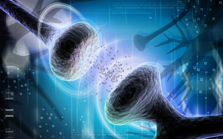
synapse
People and animals can rapidly learn to associate environmental stimuli with positive or negative outcomes, learning what to approach or avoid as they go through daily life. The amygdala plays a role in this type of emotional learning, which can be disrupted by mood disorders. In new research, Praneeth Namburi and colleagues determined that activity at the synapses in the basolateral amygdala reveals differences in the creation of fear memories and reward memories.
In animals trained with reward and fear conditioning tasks, photostimulation of neurons that then travel from the basolateral amygdala complex to the nucleus accumbens (the brain’s reward center) is positively reinforcing, while photostimulation of neurons that will travel from the basolateral amygdala complex to the centromedial nucleus of the amygdala causes aversion. There are genetic differences between the two types of neurons, including a difference in the gene for the neurotensin-1 receptor. The researchers found that neurotensin, a neuropeptide, modulates glutamate’s effect on neurons, causing some to project to the nucleus accumbens and some to project to the centromedial nucleus of the amygdala.
The researchers wrote that the results “provide a mechanistic explanation, on both a synaptic and circuit level, for how positive and negative associations can be rapidly formed, represented, and expressed within the amygdala.”
Editor’s Note: The amygdala’s creation of opposing outputs may provide clues to the mechanisms behind mania (involving the nucleus accumbens) and depression (involving the centromedial nucleus of the amygdala).
HDAC Inhibitor Facilitates Extinction of Fear Memories in the Reconsolidation Window

Unwanted recall and re-experiencing of traumatic memories is thought to be a crucial mechanism leading to the onset of post-traumatic stress disorder (PTSD). The inability to diminish (extinguish) those memories contributes to the persistence of PTSD. A new study suggests that the extinction of fear memories can be enhanced by a drug that acts epigenetically to alter the structure of DNA and subsequent gene expression.
DNA is wound around structures called histones, and chemical changes can affect how loosely or tightly the DNA is wound. Johannes Graff et al. reported in the journal Cell in 2014 that application of a histone deacetylase (HDAC) inhibitor, which keeps acetyl groups on histones, ensuring that DNA is wrapped more loosely and is easier to activate (or transcribe), helps rodents revise both new and old fear memories after they have been actively recalled.
When a memory is actively recalled, the trace of that memory in the brain becomes more amenable to revision over the proceeding five minutes to one hour (a period known as the reconsolidation window). New learning and extinction training (to get rid of the memory) lasts much longer when it takes place during the reconsolidation window than when the same procedures are performed 6 hours later (after the reconsolidation window has closed) or if the procedures are performed in the absence of active recall of the memory (when the reconsolidation window is never opened).
We have previously described the 2013 work of Xue et al. published in the journal Science, which showed that this specific procedure could yield long-lasting extinction of a patient’s craving for cocaine or heroin, and could reduce amygdala activation (as observed via functional magnetic resonance imaging) in response to an experiment that produces conditioned fear (Agren et al. Science, 2013).
Editor’s Note: This new work by Graff et al. adds another twist. Older long-term memories are more stable and less amenable to new learning than more recent (but still long-term) memories. The application of an HDAC inhibitor changes this and makes even very old memories amenable to lasting revision. The HDAC inhibitor that Graff et al. used was a specific inhibitor for HDAC type II. However, the anticonvulsant valproate (Depakote) is a potent although nonspecific HDAC inhibitor, and presumably could have the same facilitating effect as the more selective drug.
EMDR (Eye Movement Desensitization and Reprocessing), which has been widely used for the treatment of PTSD, includes active memory recall, immediately followed by an attempt to re-interpret and construct new memories of the trauma. These elements could open the reconsolidation window. However, EMDR works less well with older memories compared to more recent traumatic memories.
The Graff et al. data would suggest that adding an HDAC inhibitor such as valproate to EMDR-like work might make it more effective in revising more remote memories. Graff et al. encourage controlled clinical trials with a type II inhibitor to confirm that their findings in rodents would generalize to humans. While awaiting such validation through controlled clinical trials, it would not be surprising if clinicians started trying out the paradigm on their own using valproate.
Rats Learn Fear Conditioning From One Another

Rats who are taught to associate a light with an electric shock learn to avoid the light. This process is known as conditioned fear. New research shows that if one rat watches another rat go through fear conditioning, the observing rat will also show the effects of fear conditioning. It will also avoid the light, but only if it had previous experience with fear conditioning. It appears that rats have the ability to learn from other rats’ painful experiences.
Fear Memories Can Be Erased or Provoked in Animals

Researchers have identified neurons responsible for remembering conditioned fear in the amygdala of rodents, and can turn them on and off. At the 2013 meeting of the Society of Biological Psychiatry, Sheena A. Josselyn gave a breath-taking presentation on this process.
When animals hear a tone they have learned to associate with the imminent delivery of a shock in a given environment, they learn to avoid that environment, and they reveal their learning of the tone-shock association by freezing in place. Josselyn was able to observe that 20% of the neurons in the lateral nucleus of the amygdala were involved in this memory trace. They were revealed by their ability to increase the transcription factor CREB, which is a marker of cell activation. Using cutting-edge molecular genetic techniques, the researchers could selectively eliminate only these CREB-expressing neurons (using a new technology in which a diphtheria toxin is attached to designer receptors exclusively activated by designer drugs, or DREADDs) and consequently erase the fear memory.
The researchers could also temporarily inhibit the memory, by de-activating the memory trace cells, or induce the memory, so that the animal would freeze in a new context. Josselyn and colleagues were able to identify the memory trace for two different tones in two different populations of amygdala neurons.
The same molecular tricks with memory also worked with cocaine cues, using what is known as a conditioned place preference test. A rodent will show a preference for an environment where it received cocaine. Knocking out the selected neurons would remove the memory of the cocaine experience, erasing the place preference.
The memory for cocaine involved a subset of amygdala neurons that were also involved in the conditioned fear memory trace. Incidentally, Josselyn and her group were eventually able to show that amygdala neurons were in competition with each other as to whether they would be involved in the memory trace for conditioned fear or for the conditioned cocaine place preference.
Psychiatric Revolution: Changes in Behavior Are Associated with Dendritic Spine Shape and Number
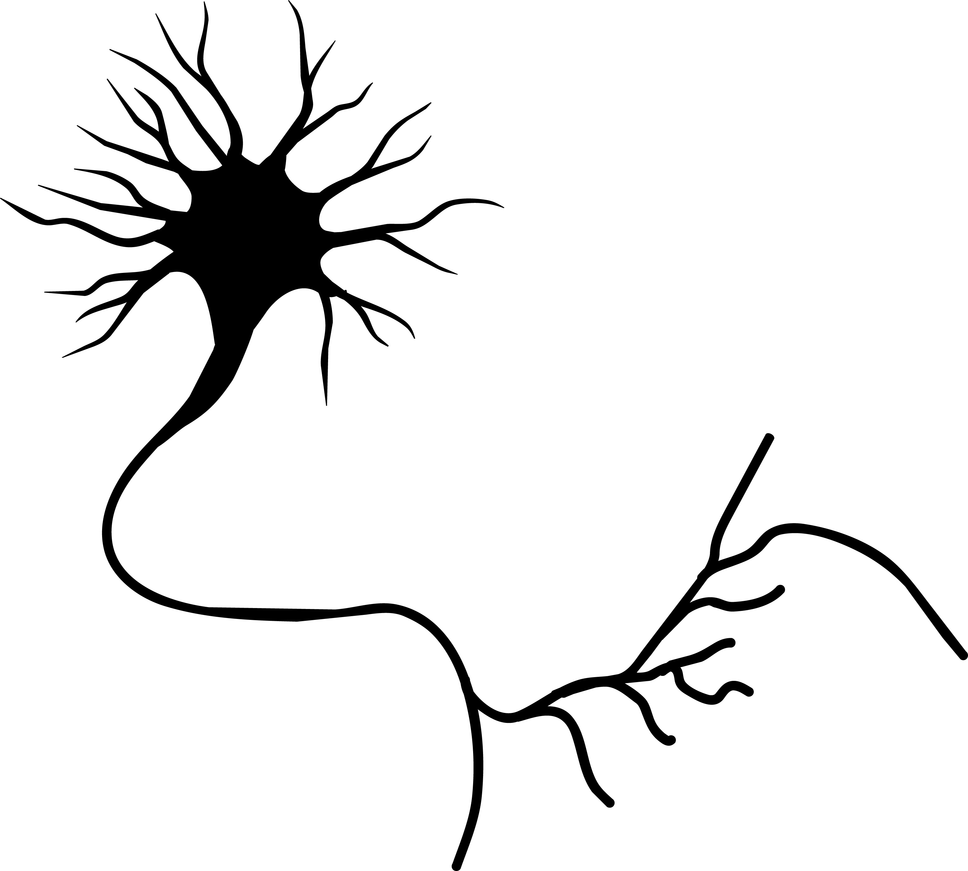
Neuron cell
New research shows that cocaine, defeat stress, the rapid-acting antidepressant ketamine, and learning and memory can change the size, shape, or number of spines on the dendrites of neurons. Dendrites conduct electrical impulses into the cell body from neighboring neurons.
Cocaine
Several researchers, including Peter Kalivas at the Medical University of South Carolina, have reported that cocaine increases the size of the spines on the dendrites of a certain kind of neurons (GABAergic medium spiny neurons) in the nucleus accumbens (the reward center in the brain). This occurs through a dopamine D1 selective mechanism. N-acetylcysteine, a drug that can be found in health food stores, decreases cocaine intake in animals and humans, and also normalizes the size of dendritic spines.
Depression
Depression in animals and humans is associated with decreases in Rac1, a protein in the dendritic spines on GABA neurons in the nucleus accumbens. Rac1 regulates actin and other molecules that alter the shape of the spines.
In an animal model of depression called defeat stress, rodents are stressed by repeatedly being placed in a larger animal’s territory. Their subsequent behavior mimics clinical depression. This kind of social defeat stress decreases Rac1 and causes spines to become thin and lose some function. Replacing Rac1 returns the spines to a more mature mushroom shape and reverses the depressive behavior of these socially defeated animals. Researcher Scott Russo has also found Rac1 deficits in the nucleus accumbens of depressed patients who committed suicide. Russo suggests that decreases in Rac1 are responsible for the manifestation of social avoidance and other depressive behaviors in the defeat stress animal model, and that finding ways to increase Rac1 in humans would be an important new target for antidepressant drug development.
Another animal model of depression called chronic intermittent stress (in which the animals are exposed to a series of unexpected stressors like sounds or mild shocks) also induces depression-like behavior and makes the dendritic spines thin and stubby. The drug ketamine, which can bring about antidepressant effects in humans in as short a time as 2 hours, rapidly reverses the depressive behavior in animals and converts the spines back to the larger, more mature mushroom-shape they typically have.
Learning and Extinction of Fear
Researcher Wenbiao Gan has reported that fear conditioning can change the number of dendritic spines. When animals hear a tone paired with an electrical shock, they begin to exhibit a fear response to the tone. In layer 5 of the prefrontal cortex, spines are eliminated when conditioned fear develops, and are reformed (near where the eliminated spines were) during extinction training, when animals hear the tones without receiving the shock and learn not to fear the tone. However, in the primary auditory cortex the changes are opposite: new spines are formed with learning, and spines are eliminated with extinction.
Editor’s Note: It appears that we have arrived at a new milestone in psychiatry. In the field of neurology, changes seen in the brains of patients with strokes or Alzheimer’s dementia have been considered “real” because cells were obviously lost or dead. Psychiatry, in comparison, has been considered a soft science because neuronal changes have been more difficult to see and illnesses were and still are called “mental.” Now that new technologies have made a deeper level of precision, observation, and analysis possible, we know that the brain’s 12 billion neurons and 4 times as many glial cells are exquisitely plastic–capable of biochemical and structural changes that can be reversed using appropriate therapeutic maneuvers.
The changes associated with abnormal behaviors, addictions, and even normal processes of learning and memory now have clearly been shown to correspond with the size, shape, and biochemistry of dendritic spines. These subtle, reproducible changes in the brain and body are amenable to therapeutic intervention, and are now even more demanding of sophisticated medical attention.
An Animal Model of Poor Judgment in Adolescence: Previous Learning Suppressed

As young mice transition into adolescence, they experience a “sensitive” period in which their context-based fear memories are temporarily suppressed. In a recent study, young animals learned to avoid an environment associated with a mild shock. Later, when they entered adolescence, this learning was temporarily forgotten or suppressed. However, when the same mice aged into adulthood, they reacquired this learned fear memory and began to again avoid the environment associated with the earlier shock. This temporary loss of fear memory differs in mice depending on their genes.
At the 2012 meeting of the Society of Biological Psychiatry, researcher Francis S. Lee reported that mice with a certain genetic variation display an impairment of this fear memory process. There are several common variants of the gene responsible for the production of brain-derived neurotrophic factor (BDNF), which protects neurons and is necessary for long-term memory. Mice with the poorer functioning variant known as Val66Met (as opposed to the better functioning Val66Val) fail to recall the earlier fear-related events not only in adolescence, but also in adulthood when the fear memory is usually retrievable again.
Editor’s Note: In mice and humans, Val66Val is the most frequently occurring allele in the population, but Val66Met is also a fairly common variation of the BDNF gene. It is this Val66Met allele that is associated with not retaining earlier learned experience about a “dangerous” environment that should be avoided.
These data suggest an intriguing explanation for some of the “wild” behavior and poor judgment to which even the smartest adolescents are prone. This kind of behavior may be based in part on the temporary forgetting in adolescence of earlier learning about which situations or environments are safe versus which ones are dangerous. Read more
Opening The Reconsolidation Window to Extinguish Fear Memories: A New Conceptual Approach to Psychotherapeutics
 Memory processes occur in several phases. Short-term memory is converted to long-term memory by a process of consolidation that requires the synthesis of new proteins. Transcription factors in the nuclei of hundreds of millions of nerve cells are activated so that specific synapses can be modified for the long term. If protein synthesis is inhibited during a period within a few hours after new learning has occurred, what was learned never gets consolidated and is essentially forgotten. It is thought that this phase of consolidation happens when a memory trace moves from short-term storage in the hippocampus to long-term storage in the cerebral cortex.
Recently a later phase of memory storage called reconsolidation has been identified. When an old memory is recalled, the reconsolidation window opens, and the memory trace becomes temporarily amenable to change. The reconsolidation window (the period during which the trace can be revised) is thought to begin five minutes after a memory is recalled and last for an hour or possibly two. New learning that takes place during the reconsolidation window can be more profound than learning that occurs without recall of the related memory or after the reconsolidation window has closed.
Consider the example of a fearful memory created when a person is attacked in a dark alley. If the person repeatedly visits the same alley without being attacked, they can eventually become less afraid of dark places. Repeated viewing of pictures of dark places can also extinguish the fear. These are typical ways in which a fear memory is extinguished. However, the original fear is subject to spontaneous recovery (the fear of dark places returns without provocation) or to reactivation (if another dangerous situation is encountered, the person may regain their fear of dark alleys).
The new findings suggest that if the extinction process (the repeated exposures to the pictures of the dark alley) takes place during the reconsolidation window after the fear memory of being attacked is recalled, the old fear can be permanently reversed (wiped clean, or re-edited such that it appears forgotten) so that it is no longer subject to spontaneous recovery or reactivation.
Editor’s Note: To accomplish extinction training within the reconsolidation window, first a person must actively recall the old memory, opening the reconsolidation window. Then, after a 5-minute delay, extinction training (e.g. new learning that the old feared place is now safe) should take place within the next hour. This process has been demonstrated in animal studies and is thought to be clinically relevant for humans in the case of phobic anxiety and post-traumatic stress disorder (PTSD). The psychotherapeutic implications of using the reconsolidation window to better ameliorate PTSD fears, avoidance, and flashbacks are enormous.
Observing the Amygdala’s Role in the Extinction of Fear Memory Traces
The amygdala is a crucial part of the learned or conditioned fear pathway. It is activated during fear conditioning and during the recall of cues associated with the fear experience. If the amygdala is removed, conditioning fear does not occur.
A new study published in Science this year by Agren et al. indicates that in humans, the amygdala-based response to conditioned fear can be completely abolished using extinction training within the memory reconsolidation window. Training that took place 10 minutes after the fear memory was activated was successful, while training that took place 6 hours later was not. Read more
