Clarifying the role of melatonin receptors in sleep
The antidepressant agomelatine (which is available in many countries, but not the US) and the anti-insomnia drug ramelteon (Rozerem) both act as agonists at melatonin M1 and M2 receptors. New research is clarifying the role of these receptors in sleep.
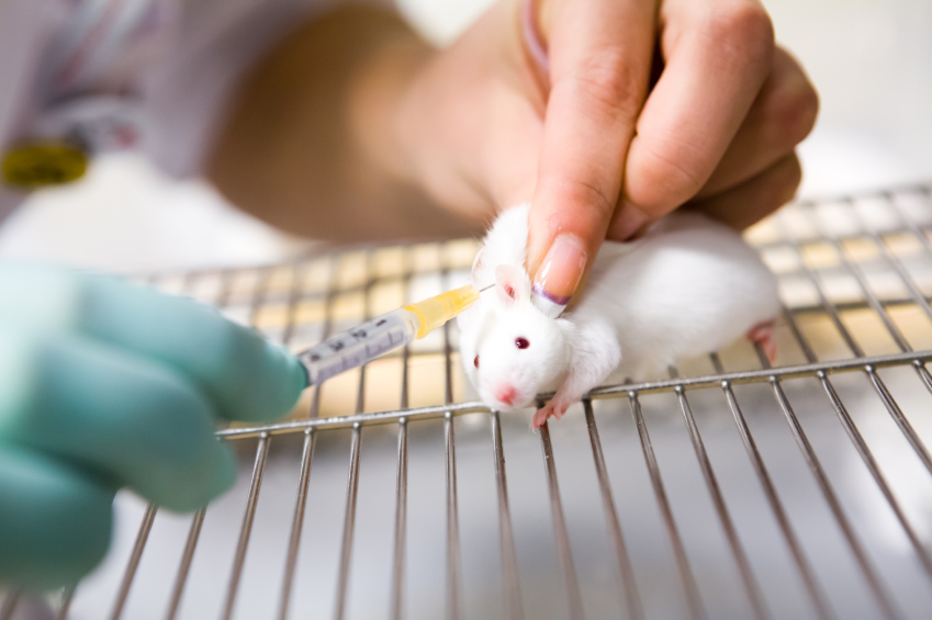
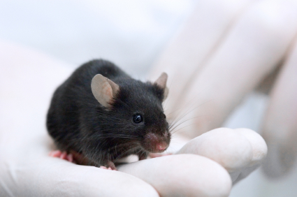
In new research from Stefano Comai et al., mice who were genetically altered to have no M1 receptor (MT1KO knockout mice) showed a decrease in rapid eye movement (REM) sleep, which is linked to dreaming, and an increase in slow wave sleep. Mice who were missing the M2 receptor (MT2KO knockout mice) showed a decrease in slow wave sleep. The effects of knocking out a particular gene like M1 or M2 end up being opposite to the effect of stimulating the corresponding receptor.
The researchers concluded that MT1 receptors are responsible for REM sleep (increasing it while decreasing slow wave sleep), and MT2 receptors are responsible for slow wave non-REM sleep.
The new information about these melatonin receptors may explain why oral melatonin supplements can make a patient fall asleep faster, but do not affect the duration of non-REM sleep. The authors suggest that targeting MT2 receptors could lead to longer sleep by increasing slow wave sleep, potentially helping patients with insomnia.
Methamphetamine Kills Dopamine Neurons in the Midbrain of Mice
Epidemiological studies have linked methamphetamine use to risk of Parkinson’s disease, and animal studies of the illicit drug have shown that it harms dopamine neurons. A 2014 study by Sara Ares-Santos et al. in the journal Neuropsychopharmacology compared the effects of repeated low or medium doses to those of a single high dose on mice. Loss of dopaminergic terminals, where dopamine is released, was greatest after three injections of 10mg/kg given at three-hour intervals, followed by three injections of 5 mg/kg at three-hour intervals, and a one-time dose of 30mg/kg. All of the dosages produced similar rates of degeneration of dopamine neurons via necrosis (cell destruction) and apoptosis (cell suicide) in the substantia nigra pars compacta (the part of the brain that degenerates in Parkinson’s disease) and the striata.
Immune Mechanisms Are Important to the Emergence of Defeat Stress–Induced Depressive Behaviors
At a recent scientific meeting, researcher Georgia E. Hodes presented evidence that in mice, the immune system may play a role in behaviors that resemble human depression. Repeated social defeat stress (when an intruder mouse is threatened by a larger mouse defending its territory) is often used as a model to study human depression. Animals repeatedly exposed to social defeat stress start to exhibit social avoidance and lose interest in sucrose. Hodes et al. determined that interleukin 6 (IL-6), an inflammatory cytokine, or signaling molecule, secreted into the blood was crucial to these behaviors. When the researchers injected mice with antibodies that block the effects of IL-6, or when they irradiated the mice’s peripheral immune system to prevent the formation of IL-6, the depressive behaviors did not emerge following defeat stress.
Editor’s Note: There are increasing data that immunological and inflammatory mechanisms play a role in human affective disorders, and these preliminary data raise the possibility that blocking some immune mechanisms more directly in humans could be a novel therapeutic approach to explore in the future.


