Neurotransmitters Can Also Function As Epigenetic Marks

The most common epigenetic marks involve methylation of DNA (which usually inhibits gene transcription) and the acetylation and methylation of histones. Acetylation opens or loosens the winding of DNA around the histones and facilitates transcription, while methylation of histones leaves the DNA tightly wound and inhibits transcriptional activation.
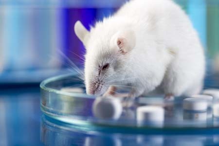
Researcher Ashley E. Lepack and colleagues have identified a surprising type of epigenetic mechanism involving neurotransmitters. They report in a 2020 article in the journal Science that neurotransmitters such as serotonin and dopamine can act as epigenetic marks. Dopamine can bind to histone H3, a process called called dopaminylation (H3Q5dop). In rats undergoing withdrawal from cocaine, Lepack and colleagues found increased levels of H3Q5dop in dopamine neurons in a part of the midbrain called the ventral tegmental area (VTA), a part of the brain’s reward system. When the investigators reduced H3Q5dop, this decreased dopamine release in the reward area of the brain (the nucleus accumbens) and reduced cocaine seeking. Thus, dopamine can be both an important transmitter conveying messages between neurons and a chemical mark on histones that alters DNA binding and transcriptional regulation.
Researcher Jean-Antoine Girault provided commentary on the article by Lepack and colleagues, writing that “[t]he use of the same monoamine molecule as a neurotransmitter and a histone modification in the same cells illustrates that evolution proceeds by molecular tinkering, using available odds and ends to make innovations.”
Editor’s Note: Epigenetic marks may remain stable and influence behavior over long periods of time. They are involved in the increased reactivity or sensitization to repeated doses of cocaine through DNA methylation. Such sensitization can last over a period of months or longer. If the methylation inhibitor zebularine is given, animals fail to show sensitization. Now a newly identified epigenetic process, dopaminylation, is found to alter histones and is associated with long-term changes in cocaine-seeking.
The clinical message for a potential cocaine user is ominous. Cocaine not only creates a short-term “high,” but its repeated use rewires the brain not only at the level of changes in neurotransmitter release and receptor sensitivity, but also at the genetic and epigenetic level, changes that could persist indefinitely.
The sensitization to motor hyperactivity and euphoria that occur with cocaine use can progress to paranoia and panic attacks and eventually even seizures (through a process known as kindling).
The dopaminylation of histones in the VTA could lead to persistent increases in drug craving and addiction that may not be easily overcome. Thus, the appealing short-term effects of cocaine can spiral into increasingly adverse behaviors and drug-seeking can become all consuming. While these adversities do not emerge for everyone, the best way to ensure that they do not is to avoid cocaine from the start.
Manic episodes that include a feeling of invincibility, increased social contacts, and what the DSM-5 describes as “excessive involvement in pleasurable activities that have a high potential for painful consequences” are a time that many are at risk for acquiring a substance problem. For the adolescent who has had a manic episode, ongoing counseling about avoiding developing this type of additional long-term, difficult-to-treatment psychiatric illness could be lifesaving. Describing the epigenetic consequences of substance use may or may not be helpful, but may be worth a try.
In Rats, Weight-Loss Drug Lorcaserin Reduces Opiate Use
The serotonin 5HT-2c agonist drug lorcaserin (Belviq) was approved by the US Food and Drug Administration in 2012 for the treatment of obesity and weight-related conditions (such as high blood pressure, type 2 diabetes, or high cholesterol) in adults. A 2017 article by researcher Harshini Neelakantan and colleagues in the journal ACS Chemical Neuroscience reports that in rats, lorcaserin may also reduce opiate use.
The rats had been self-administering the opiate oxycodone. After receiving lorcaserin, the rats were less likely to consume oxycodone and less likely to seek it out. The rats were also less responsive to cues that had previously led them to consume oxycodone, such as lights or sounds that occurred when oxycodone was available.
Serotonin 5HT-2c receptors both regulate psychostimulant reward in the brain and play a role in reactivity to cues like the lights and sounds the rats associated with oxycodone. Lorcaserin’s effect on these serotonin receptors explains how it could reduce the rats’ drug use.
Clinical trials are expected to examine whether lorcaserin can reduce opiate use in humans in addition to assisting with weight loss.
In Rats, Dad’s Cocaine Use Affects Son’s Spatial Memory
Evidence is mounting that certain behaviors by parents can leave marks on their sperm or eggs that are passed on to their offspring in a process called epigenetics. In a recent study by researcher Mathieu Wimmer and colleagues, male rats that were exposed to cocaine for 60 days (the time it takes for sperm to develop fully) had male offspring who showed diminished short- and long-term spatial memory compared to the offspring of male rats that were not exposed to cocaine. Female offspring were not affected in this way.
The spatial tasks the offspring rats completed depended heavily on the hippocampus. Wimmer and colleagues believe that cocaine use in the fathers decreased the amount of a brain chemical called d-serine in the offspring. D-serine plays a role in memory formation and the brain’s ability to form synaptic connections. Injecting the offspring of rats who were exposed to cocaine with d-serine before the spatial memory tasks normalized the rats’ performance.
Marijuana Use Worsens PTSD Symptoms in Veterans
A 2015 study by Samuel T. Wilkinson and colleagues in the Journal of Clinical Psychiatry reports that among war veterans who completed a special treatment program for post-traumatic stress disorder, those who continued or began using marijuana after treatment had more severe PTSD symptoms, were more violent, and used drugs and alcohol more often. Those who stopped using marijuana or never used it had the lowest levels of PTSD symptoms in the study.
Editor’s Note: Scientific information about marijuana is almost never reported in the media. Evidence of the adverse effects of heavy marijuana use are robust and consistent.
Some of these include:
- A doubling of the risk of psychosis compared to non-users. People with a common variation in the enzyme COMT, which metabolizes dopamine, have an even higher rate of psychosis.
- An increased risk of bipolar disorder onset.
- A worse course of bipolar disorder.
- An increased risk of schizophrenia.
- Memory deficits that remain even after marijuana use has ceased.
- Loss of motivation (exactly what someone with depression doesn’t need).
- Anatomical changes in brain structures.
- A worse course of PTSD and increased violence in those with PTSD.
Bottom line: Those who say marijuana is benign may be ill-informed. People with mood disorders, proneness to paranoia, or PTSD should stay away from marijuana.
Methamphetamine Kills Dopamine Neurons in the Midbrain of Mice
Epidemiological studies have linked methamphetamine use to risk of Parkinson’s disease, and animal studies of the illicit drug have shown that it harms dopamine neurons. A 2014 study by Sara Ares-Santos et al. in the journal Neuropsychopharmacology compared the effects of repeated low or medium doses to those of a single high dose on mice. Loss of dopaminergic terminals, where dopamine is released, was greatest after three injections of 10mg/kg given at three-hour intervals, followed by three injections of 5 mg/kg at three-hour intervals, and a one-time dose of 30mg/kg. All of the dosages produced similar rates of degeneration of dopamine neurons via necrosis (cell destruction) and apoptosis (cell suicide) in the substantia nigra pars compacta (the part of the brain that degenerates in Parkinson’s disease) and the striatum.


