Heart Attacks, Surgery Lead to Memory Impairment in Mice
Events like surgery or heart attacks that cause inflammation can lead to cognitive deficits or depression for months or years afterward, even though the direct effects of inflammation wear off within weeks. In a recent study, Natalie Tronson and colleagues subjected mice to surgical heart attack, sham surgery, or no operation, and observed how well they absorbed new learning eight weeks later.
Both male and female mice had impairments in fear learning following surgical heart attacks. Female mice that received sham surgery also showed deficits in fear learning. When the researchers dissected the mice, analyzing their blood and hippocampi after the eight-week period, inflammatory cytokine measures had normalized as expected, but the researchers found other abnormalities.
Intracellular signaling was dysregulated, and there had been epigenetic changes in cells of the hippocampus. (Epigenetic changes refer to those that change the structure of DNA, such as how tightly it is wound, rather than its sequence. For example, the addition of acetyl groups to DNA or the histones around which it is wound.) The researchers observed increased histone acetylation and phospho-acetylation following the heart attacks.
The researchers concluded that a systemic inflammatory event, such as heart attack or surgery, can cause long-term memory impairment and changes in mood through epigenetic mechanisms. They compared the findings to those of other studies in which normal aging and memory-impairing treatments such as chemotherapy had also been associated with increases in histone acetylation or decreases in histone deacetylase activity.
Early Life Stressors Lead to Lifetime Increase in Inflammation in Mice
Stressors in early life can contribute to the risk of developing mood disorders. Given that many treatments for mood disorders work by blocking the serotonin 5-HT transporter, Nicole Baganz and colleagues designed a study to see whether an early life stressor, in this case maternal separation, would affect immune processes that in turn affect serotonin signaling.
In this study as in many before it, mice that were removed from their mothers exhibited behaviors that resembled human anxiety and depression. They were also found to have elevated messenger RNA for several inflammatory cytokines (including IL-1beta and IL-6) in their brain and blood. Mice that had a gene for the interleukin-1 receptor (IL-1R) removed exhibited neither the depressive behavioral effects nor the changes in cytokine levels following maternal separation, showing that the IL-1R gene plays a necessary role in the signaling process that leads to this type of depression. Levels of the stress hormone corticosterone in the blood did not differ in the mice with and without the IL-1R gene.
The researchers concluded that early life stressors can cause lifelong changes in inflammatory cytokine levels in mice.
Young Rats That Witness Maternal Abuse Show Depression-Like Behavior in Adulthood
Rodents that are subjected to social defeat (being overpowered by a bigger, more aggressive animal) develop a syndrome that resembles human depression—they avoid social interaction, lose interest in sucrose, and do less exploring of new places or other animals. A recent finding showed that even witnessing the social defeat of a peer was enough to bring about the depressive behaviors. The same researchers, led by Samina Salim, recently found that young rats (aged 21–27 days) that witnessed their mother go through the trauma of social defeat showed depression-like behavior themselves as adults (at age 60 days).
The rats saw their mothers defeated by the larger rat every day for seven days. As adults, those who witnessed this abuse exhibited depression-like behavior compared to rats of the same age and gender that had not witnessed abuse. The depressive rats gave up more quickly on a test of forced swimming. Male rats showed great depression-like behavior than female rats.
It has been estimated by the American Psychological Association that 15.5 million children in the US witness physical or emotional abuse of a parent (usually their mother). Children who witness domestic violence often show symptoms of post-traumatic stress disorder (PTSD). This rodent research may lead to a better understanding of the consequences of witnessing trauma in childhood, and potential treatments that could help.
Editor’s Note: These data show that rats have something like empathy, and that the psychological aspects of stress (including verbal abuse in humans and witnessing another’s abuse in rodents) may have profound and lasting consequences on behavior.
RTMS Can Increase Amygdala Connectivity
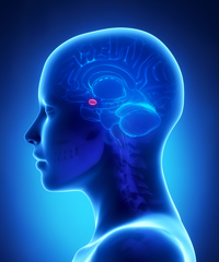
 Regulation of the amygdala (the brain’s emotional center), particularly through its interaction with the ventral anterior cingulate cortex, has been implicated in the experience of fear in animals, and anxiety and depression in humans. Connectivity between the two structures is critical for emotion modulation. Repeated transcranial magnetic stimulation (rTMS) is a method of stimulating outer regions of the brain with magnets. Researchers Desmond Oathes and Amit Etkin are investigating whether rTMS can also be used to influence these deeper brain areas, or their interaction with each other.
The researchers’ study used single-pulse probe TMS delivered at a rate of 0.4 Hz at 120% of each participant’s motor threshold, targeted at the anterior or posterior medial frontal gyrus on either side of the brain. The researchers also used functional magnetic resonance imaging (fMRI) of the whole brain to observe connectivity between different sections.
RTMS to the right side of the medial frontal gyrus increased connectivity between the amygdala and the ventral anterior cingulate cortex more than stimulation to the left side. Stimulation of the posterior portion of the medial frontal gyrus increased connectivity more than stimulation of the anterior portion.
Editor’s Note: These data indicate that rTMS can alter brain activity in these deeper regions and can influence inter-regional connectivity. This is important because abnormalities in the connectivity of brain regions have increasingly been found in patients with mood disorders. Oathes and Etkin hope that these findings can be applied to others and that rTMS can be used to correct patterns of regional connectivity in the brain in order to improve emotion regulation.
ECT versus Drug Therapy for Bipolar Depression
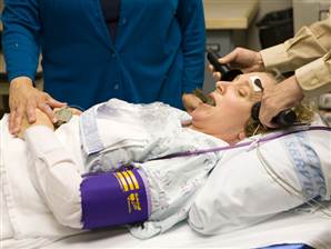
 Electroconvulsive therapy is often considered a primary treatment option for patients with severe bipolar disorder that has resisted pharmacological treatment. Researcher Helle K. Schoeyen and colleagues recently published the first randomized controlled trial comparing ECT (in this case right unilateral brief pulse ECT) with algorithm-based pharmacological treatment in 76 patients with treatment-resistant bipolar depression.
The response rate was significantly higher in the ECT group than in the patients who received drug treatment (73.9% versus 35.0%). However, the two treatment groups had similarly low remission rates (34.8% for ECT and 30.0% for pharmacological treatment).
The algorithm-based pharmacological treatment used in the study was based on a sequence of treatments endorsed by researchers Frederick K. Goodwin and Kay Redfield Jamison in their 2007 book Manic-Depressive Illness. A selected treatment was chosen for each participant based on his or her medical history. If the first treatment was ineffective or intolerable, the patient would be switched to the next treatment option. Antipsychotics, antidepressants, anxiety-reducing drugs, and hypnotics were some of the other treatments included in the algorithms.
Patients in the study had previously showed a lack of response to at least two different antidepressants and/or mood stabilizers with documented efficacy in bipolar disorder (lithium, lamotrigine, quetiapine, or olanzapine) in adequate doses for a period of 6 weeks (or until they quit because of side effects).
Editor’s Note: Even when ECT is effective, there is the issue of how to maintain that good response. We previously reported that in a 2013 study by Axel Nordenskjöld et al. in the Journal of ECT, intensive followup treatment with right unilateral brief pulse ECT combined with pharmacotherapy was more effective than pharmacology alone at preventing relapses. Patients who improved after an acute series of ECT (three times/week) then received weekly ECT for six weeks and every two weeks thereafter, totaling 29 ECT treatments in one year.
Other studies of more intermittent continuation ECT have not proved more effective than medication. Thus high intensity right unilateral brief pulse ECT is one option for extending the effects of successful ECT.


