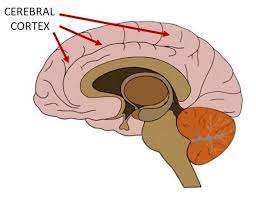
Surface Area of Cortex Is Reduced After Multiple Manic Episodes

In a 2020 article in the journal Psychiatric Research: Neuroimaging, researcher Rashmin Achalia and colleagues described a study of structural magnetic resonance imaging (MRI) that compared 30 people with bipolar I disorder who had had one or several episodes of mania to healthy volunteers. Compared to the healthy volunteers, people with bipolar disorder had “significantly lower surface area in bilateral cuneus, right postcentral gyrus, and rostral middle frontal gyri; and lower cortical volume in the left middle temporal gyrus, right postcentral gyrus, and right cuneus.”
The surface area of the cortex in patients with bipolar I disorder who had had a single episode of mania resembled that of the healthy volunteers, while those who had had multiple manic episodes had less cortical surface area.
The data suggest that compared to healthy volunteers, people with bipolar disorder have major losses in brain surface area after multiple episodes that are not seen in first episode patients. In addition, the researchers found that both the number of episodes and the duration of illness was correlated with the degree of deficit in the thickness in the left superior frontal gyrus. These decreases in brain measures occurred after an average of only 5.6 years of illness.
Editor’s Note: These data once again emphasize the importance of preventing illness recurrence from the outset, meaning after the first episode. Preventing episodes may prevent the loss of brain surface and thickness.
Clinical data has also shown that multiple episodes are associated with personal pain and distress, dysfunction, social and economic losses, cognitive deficits, treatment resistance, and multiple medical and psychiatric comorbidities. These and other data indicate that treatment after a first episode must be more intensive, multimodal, and continuous and include expert psychopharmacological and psychosocial support, as well as family education and support. Intensive treatment like this can be life-saving. The current study also supports the mantra we have espoused: prevent episodes, protect the brain and the person.
In Animal Model, Long-Term THC Exposure Interferes with Cortical Control of the Nucleus Accumbens

In an article in the journal Biological Psychiatry, researchers Eun-Kyung Hwang and Carl R. Lupica reported that in rats, long-term use of THC (tetrahydrocannabinol) weakens input from the cortex to the reward area of the brain, the nucleus accumbens (NAc). Long-term THC use also strengthens connections to the NAc from emotional control (limbic) regions, such as the basolateral amygdala and ventral hippocampus. Hwang and Lupica reason that this shift from cortical control of the NAc to limbic control likely contributes to the cognitive and psychiatric symptoms associated with cannabis use.
Editor’s Note: Street marijuana largely contains THC rather than CBD, the beneficial, anxiety-reducing component of cannabis. Cannabis products are being decriminalized, but it is important to remember that those with THC are linked to cannabis use disorder and increased susceptibility to psychiatric illness. Patients with bipolar disorder who use marijuana also have a more adverse course of illness than those who do not use it.
Marijuana May Speed Cortical Loss in Boys at Risk for Schizophrenia
In boys, a decrease in the thickness of the cortex is a part of normal maturation. However, according to a recent study, this process is sped up in boys at high risk for schizophrenia when they use marijuana before the age of 16.
Early use of marijuana has been linked to subsequent development of schizophrenia. Schizophrenia begins about 5 years earlier in males than in females, and the male brain goes through more structural changes during adolescence.
A 2015 article by Tomáš Paus in the journal JAMA Psychiatry incorporated data from three studies, which took place in parts of Canada and England and eight European cities. The studies all included magnetic resonance imaging (MRI) scans of the participants, a measure of their genetic risk of developing schizophrenia, and questions about their past marijuana use. In boys at high risk for schizophrenia based on their genetic profile, cortical thickness dropped more among the ones who used high amounts of marijuana before the age of 16 compared to those who did not.
Paus hypothesizes that the development of schizophrenia is a “two-hit process.” People who develop schizophrenia may have an early risk factor, such as their genetic profile or a problem that occurs in utero, and a later stressor such as drug use in adolescence.


